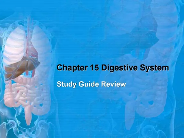
Chapter 15 The Digestive System
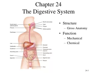
Chapter 15 The Digestive System. DIGESTIVE SYSTEM (FIGURE 15-1). Irregular tube called alimentary canal or gastrointestinal (GI) tract Food must first be digested, then absorbed, and later metabolized. WALL OF THE DIGESTIVE TRACT (FIGURE 15-2).

Chapter 15 The Digestive System
E N D
Presentation Transcript
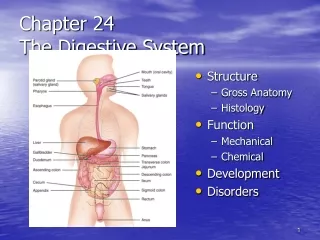
DIGESTIVE SYSTEM (FIGURE 15-1) • Irregular tube called alimentary canal or gastrointestinal (GI) tract • Food must first be digested, then absorbed, and later metabolized
WALL OF THE DIGESTIVE TRACT (FIGURE 15-2) • Digestive tract described as tube that extends from mouth to anus • Wall of the digestive tube is formed by four layers of tissue: • Mucosa—mucous epithelium • Submucosa—connective tissue • Muscularis—two layers of smooth muscle • Serosa—serous membrane that covers the outside of abdominal organs; it attaches the digestive tract to the wall of the abdominopelvic cavity by forming folds called mesenteries
MOUTH • Roof—formed by hard palate (parts of maxillary and palatine bones) and soft palate, an arch-shaped muscle separating mouth from pharynx; uvula, a downward projection of soft palate (Figure 15-4) • Floor—formed by tongue and its muscles; papillae, small elevations on mucosa of tongue; taste buds, found in many papillae; lingual frenulum, fold of mucous membrane that helps anchor tongue to floor of mouth (Figure 15-4) • Typical tooth (Figure 15-5) • Three main parts—crown, neck, and root • Enamel, which covers the crown, is hardest tissue in body
MOUTH • Types of teeth—incisors, cuspids, bicuspids, and tricuspids • Twenty teeth in temporary set; average age for cutting first tooth about 6 months; set complete at about 2 years of age • Thirty-two teeth in permanent set; 6 years about average age for starting to cut first permanent tooth; set complete usually between ages of 17 and 24 years (Figure 15-6)
SALIVARY GLANDS (FIGURE 15-7) • Parotid glands—largest salivary glands • Submandibular glands—open into mouth on either side of frenulum • Sublingual glands—open into floor of mouth
PHARYNX • Subdivided into three anatomical components: • Nasopharynx • Oropharynx • Laryngopharynx
ESOPHAGUS • Connects pharynx to stomach • Dynamic passageway for food
STOMACH (Figure 15-8) • Size—expands after large meal; about size of large sausage when empty • Food enters stomach through gastroesophageal (cardiac) sphincter • Pyloric sphincter muscle closes opening between pylorus (lower part of stomach) and duodenum • Wall—many smooth muscle fibers; contractions produce churning movements (peristalsis) • Lining—mucous membrane; many microscopic glands that secrete gastric juice and hydrochloric acid into stomach; mucous membrane lies in folds (rugae) when stomach is empty
SMALL INTESTINE (FIGURE 15-9) • Size—about 7 meters (20 feet) long but only 2 cm or so in diameter • Divisions • Duodenum • Jejunum • Ileum
SMALL INTESTINE • Wall—contains smooth muscle fibers that contract to produce peristalsis • Lining—mucous membrane; many microscopic glands (intestinal glands) secrete intestinal juice; villi (microscopic finger-shaped projections from surface of mucosa into intestinal cavity) contain blood and lymph capillaries
LIVER AND GALLBLADDER • Size and location—liver is largest gland; fills upper right section of abdominal cavity and extends over into left side • Liver secretes bile • Ducts (Figure 15-10) • Hepatic—drains bile from liver • Cystic—duct by which bile enters and leaves gallbladder • Common bile—formed by union of hepatic and cystic ducts; drains bile from hepatic or cystic ducts into duodenum • Gallbladder • Location—undersurface of the liver • Function—concentrates and stores bile produced in the liver
PANCREAS • Exocrine gland that lies behind stomach • Functions • Pancreatic cells secrete pancreatic juice (most important digestive juice) into pancreatic ducts; main duct empties into duodenum • Pancreatic islets (of Langerhans)—cells not connected with pancreatic ducts; secrete hormones glucagon and insulin into the blood
LARGE INTESTINE (FIGURE 15-12) • Divisions • Cecum • Colon—ascending, transverse, descending, and sigmoid • Rectum • Food enters through ileocecal valve; external opening called anus • Wall—contains smooth muscle fibers that contract to produce churning, peristalsis, and defecation • Lining—mucous membrane
APPENDIX • Blind tube off cecum • No important digestive functions in humans
PERITONEUM (FIGURE 15-14) • Definitions—peritoneum, serous membrane lining abdominal cavity and covering abdominal organs; parietal layer of peritoneum lines abdominal cavity; visceral layer of peritoneum covers abdominal organs; peritoneal space lies between parietal and visceral layers • Extensions—largest are the mesentery and greater omentum • Mesentery is extension of parietal peritoneum, which attaches most of small intestine to posterior abdominal wall • Greater omentum, or “lace apron,” hangs down from lower edge of stomach and transverse colon over intestines • X-ray studies of the GI tract—radiopaque contrast medium used to help visualize structures in study images
DIGESTION (TABLE 15-2) • Definition—transforms foods into substances that can be absorbed and used by cells • Mechanical digestion—chewing (mastication), swallowing (deglutition), and peristalsis break food into tiny particles, mix them well with digestive juices, and move them along the digestive tract • Chemical digestion—breaks up large food molecules into compounds that have smaller molecules; brought about by digestive enzymes (Figure 15-15) • Enzymes and chemical digestion • Enzymes are specialized protein molecules that act as catalysts • Breakdown process called hydrolysis
DIGESTION • Carbohydrate digestion—mainly in small intestine • Pancreatic amylase—breaks polysaccharides down to disaccharides • Intestinal juice enzymes • Maltase—changes maltose to glucose • Sucrase—changes sucrose to glucose • Lactase—changes lactose to glucose
DIGESTION • Protein digestion—starts in stomach; completed in small intestine • Gastric juice enzyme pepsin partially digests proteins • Pancreatic enzyme, trypsin, continues digestion of proteins • Intestinal enzymes, peptidases, complete digestion of partially digested proteins and convert them to amino acids • Fat digestion • Bile contains no enzymes but emulsifies fats (breaks fat droplets into very small droplets) • Pancreatic lipase changes emulsified fats to fatty acids and glycerol in small intestine
ABSORPTION • Definition—process by which digested food moves from intestine into blood or lymph • Foods and most water minerals and vitamins are absorbed from small intestine; some water and vitamin K also absorbed from large intestine • Surface area absorption • Structural adaptations increase absorptive surface area • Fractal geometry—study of fragmented geometric irregular shapes such as those in lining of intestine