Download
1 / 42
420 likes | 643 Views
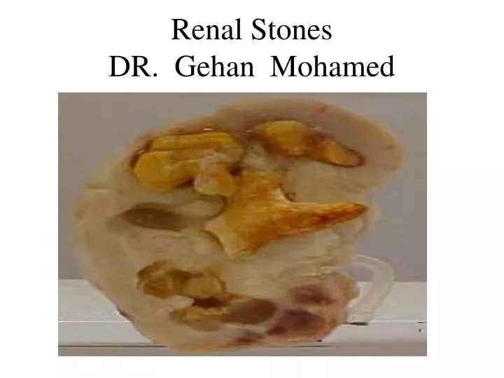
Renal Stones DR. Gehan Mohamed. Learning objectives. 1- define renal stones and know the alternative names. 2- understand the causes and risk factors for the formation of nephrolithiasis. 3- mention the Inhibitors of Spontaneous Crystallization in urine.

E N D
Learning objectives • 1- define renal stones and know the alternative names. • 2- understand the causes and risk factors for the formation of nephrolithiasis. • 3- mention the Inhibitors of Spontaneous Crystallization in urine. • 4- list the different types of renal stones • 5- understand cause , site of the renal colic. • 6- mention the important investigations needed for diagnosis of nephrolithiasis. • 7- list complications and methods of treatment for renal stones.
How is Urine formed?1- blood filteration through glomeruli2- tubular reabsorption3- tubular secretion
Kidney Stones • Definition :are hard deposits of mineral and acid salts on the inner surfaces of the kidneys • Alternative names include: • Renal Lithiasis • Nephrolithiasis (Kidney Stone Disease) • Renal Calculi • Stones are classified by : - Their location in the urinary system - Their composition of crystals. - Their size : either small or large.
Inhibitors of Spontaneous Crystallization • Normal urine contains chelating agents(i.e agents have ability to make bonds with another substances) such as citrate that inhibit the nucleation, growth, and aggregation of calcium-containing crystals. • Other endogenous inhibitors include calgranulin (an S-100 calcium binding protein), Tamm-Horsfall protein
Causes of Stone formation 1-Supersaturation of urine either due to Dehydration or due to increase level of minerals in urine such as (Ca,oxalate) ,uric acid lead to Crystal aggregation 2- Bacterial Infection: - E.Coli infection increases protein matrix content in urine - Proteus makes urine alkaline as it split urea into amonia making alkaline urine so fascilitate deposition of magnesium ammonium phosphate crystals (struvite stone). 3-Imbalance of pH in urine • Acidic: Uric acid,calcium oxalate Stones • Alkaline: Calcium phosphate Stones 4-Anatomic Abnormalities as Pelviureteric junction stenosis leading to urine stasis.
Risk Factors for renal stones • 1-family history: is a factor in certain forms of stone disease such as cystinuria which is an inherited metabolic disorder. • 2- Occupation: activities that keep people out in hot weather and increased sweating as in construction work, farming . • 3- Diet: - Low fluid intake lead to supersaturation of urine - high dietary intake of: - animal protein produce purine which change to uric acid - refined sugars, fructose - grapefruit juice rich in vitamen C (ascorbate) which have the ability to change to oxalate. • 4- drugs as diuretics can cause dehydration.
Risk Factors • 5- crohn’sdisease are associated with steattorhea, The fatty acids in the gut bind with intraluminal calcium leaving the oxalate to be absorbed by blood. net effect is hyperoxaluria. • 6- HYPERCALCAEMIA & HYPERCALCIURIA either due to : - some diseases associated with hypercalcemia as hyperparathyroidism,sarcoidosis ,hyperthyroidism,myeloma,Vit. D Intoxication,Metastatic Malignant Neoplasms. - while excessive supplemental calcium in diet do not appear to cause kidney stones and may actually protect against their development.This is perhaps related to the role of oral calcium in binding ingested oxalate in the gastrointestinal tract and prevent its reabsorption by blood so prevent hyperoxaluria and stone formation.
structure of Renal Stones • all urinary stones are composed of : • crystalline material 98% which have a very limited solubility in urine . • mucoprotein 2% (matrix) • N.B The stone with branches formed within calyxes and pelvis is termed stag horn-like or coral-like stone
Stag horn stone : is the stone with branches formed within calyxes and pelvis is termed stag horn-like or coral-like stone
Types of Renal Stones (cont.) 1- calcium oxalate (75%) need acidic urine. 2- calcium hydroxyl phosphate (15%) need alkaline urine. 3- struvite magnesium ammonium phosphate 10% is found mostly in women because of their increase chance of having chronic pyelonephritis. 4- Uric acid stone (5% ): - occur in acidic urine with PH below 7 leading to crystal formation. - common in patients with gout,high protein diet ,chemotherapy treatment. - It is the only type that is truly radiolucent (can not be seen on a plain abdominal film. 5- Cystine stones ( 1%) of stones due to genetic abnormality in its metabolism leading to its leak through the kidneys and into the urine to form crystals. .
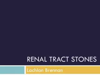
Stained and unstained microscopic image of a calcium oxalate stone in urine
Commonest Site of Stone Development Question: Where in the Urinary Tract does urine reach its maximal concentration and can get “stuck” in the lumen? Answer: at the collecting duct in the renal papilla serves as the “uterus” for stone formation. It is here that urine has achieved its maximum concentration and hence is most likely to be supersaturated. Clumps of crystals become impacted in the opening of a collecting duct so that some of the crystals are now exposed to the urine in that calyx that comes from other collecting ducts
Excretion of urine from body is essential to get rid of excess body fluids, excess electrolytes and waste products
Diagnosis of renal calculi • Clinical pictures. • urine analysis • radiographic studies
Asymptomatic if the stone donot cause any obstruction. Severe flank pain :colicky in nature (comes and goes in spasmodic waves). Pain in the back occurs when calculi produce an obstruction in the kidney or pain caused by peristaltic contractions of the ureter as it attempts to expel the stone flank pain referred to genitalia nausea, vomiting may mislead Microhematuria if stone have rough surface. Clinical Features
investigations Urine analysis : hematuria, infection,type of crystals. X - Ray : most of renal stones are radiopaque due to calcification except uric acid stones are radiolucent so not seen. Intravenous urography(IVU) will show delayed function, hydronephrosis and ureteral dilatation to point of stone Ultrasound can show hydronephrosis, stones.
Struvite crystals found on microscopic examination of the urine
Complications of renal stones 1- Calcularpyelonephritis 2-obstructive uropathy with Reflux nephrosis in the form of : - Hydronephrosis :- dilatation of renal pelvis ,calyces with atrophy of renal parenchyma due to lower obstruction . Then Hydronephrosis lead to 2ry infection due to stasis of urine producing Pyonephrosis So Pyonephrosis = Hydronephrosis (dilatation)+ infection (pus) 3-Kidney damage, scarring leading to Postrenal azotemia, renal failure .
Adequate water intake to reach urine level per day about 2 litres as Drinking enough water helps keep urine diluted and flushes away materials that might form stones that is the most important thing a person can do to prevent kidney stones. Increase citrate intake present in lemon and orange juice . but citrus drinks may be helpful in preventing calcium oxalate stones and uric acid stones but they might be harmful for people who form calcium phosphate stones. limit vitamen C,sodium ,animal protein,excess flouride in water. Reducing salt intake is preferred to reducing calcium intake because Salt is made up of sodium and chloride. The sodium in salt, when excreted by the kidneys, causes more calcium to be excreted into the urine. High concentrations of calcium in the urine combine with oxalate and phosphorus to form stones. Medical Treatment for renal stones
Limit oxalate intake present in nuts,spinach, strawberries, nuts, dark chocolate, coffee and tea. • Change PH of urine to increase solubilty of the solutes. • Antibiotic intake with struvite type
Surgical Treatment Surgery is usually needed if: • The stone is too large to pass on its own • The stone is growing • The stone is blocking urine flow and causing an infection or kidney damage • The pain cannot be controlled
treatment for renal stones • 1- ESWL :Extracorporeal shock-wave lithotripsy is used with stones smaller than a half an inch . It uses sound or shock waves to break up stones. Then, the stones leave the body in the urine. • 2- Percutaneous nephrolithotomy using endoscopeis used for large stones. The stone is removed with tube that is inserted into the kidney through a small surgical cut. • 3- Ureteroscopymay be used for stones in the lower urinary tract. • 4- open surgery (nephrolithotomy) may be needed if other methods do not work or are not possible.