
Microbial Cell Structure and Functions
E N D
Presentation Transcript
Chapter 3 Microbial Cell Structure
According to the cell theory of life, a single cell is the smallest unit of life and is capable of carrying out all of the basic processes of a living organism. 5 important characteristics of all living organisms - ability to reproduce - ability to take in food, produce energy and grow - ability to excrete wastes - ability to respond to the environment - susceptible to mutation
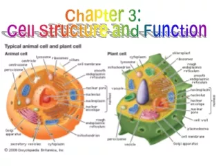
Prokaryotic vs. Eukaryotic Cells Algae 80S ribosomes Cell Wall simple – cellulose Cell Membrane phospholipids and sterols embedded proteins DNA several linear chromosomes billion base pairs mRNA spliced before translation Endoplasmic Reticulum Nuclear Membrane Organelles Mitochondria and Chloroplasts Complex Flagella sheathed, several microtubules whipping motion
How big are typical microorganisms like protozoa, bacteria and viruses? Viruses 25 – 400 nm (.025 - .400 mm) Bacteria 0.2 – 10 mm Protozoa 1 – 150 mm There is a lot of variation in the sizes of different types of microorganisms but as a general rule protozoa are bigger than bacteria and bacteria are bigger than viruses. There are of course exceptionally large bacteria and exceptionally small protozoa so one can find cases of a given bacterium that is bigger than a given protozoa.
Envelope Cell Membrane – diffusion barrier, always present Cell Wall – usually present, maintains cell shape, protects from osmotic lysis by preventing excessive swelling Glycocalyx – external layer of slime, protects bacterium from host immune system, freezing and dessication helps bacteria to stick together
Functions of the cytoplasmic membrane (cell membrane) The cytoplasmic membrane is an absolutely essential structure. All living cells are bounded by a cytoplasmic membrane. The cell membrane is a semipermiable barrier. It allows some substances to diffuse into and out of the cell but it prevents other substances from entering or leaving the cell. The cytoplasmic membrane is the structure that defines the border between the living matter within the cell and nonliving matter that is outside. In bacteria the enzymes and electron carriers of the respiratory chain are embedded in the cytoplasmic membrane. (In eukaryotic cells the respiratory enzymes are found in the inner mitochondrial membranes. Bacterial cells don't have mitochondria.)
Cytoplasmic Membrane Composition The bacterial cytoplasmic membrane is composed of a lipid bilayer that has protein molecules embedded within it. By mass a typical bacterial cytoplasmic membrane is about 50% protein and 50% lipid. The lipids found in bacterial cell membranes are phospholipids. See pages 70 – 74 of your textbook for more information about cell membranes.
Cytoplasmic Membrane Which one of the following statements about cytoplasmic membranes is FALSE? A. Virus particles may or may not have an envelope but they are NOT surrounded by an active cytoplasmic membrane. B. The cytoplasmic membrane helps to regulate diffusion of molecules into and out of the cell. C. The shape of a bacterial cell is determined by the structure of the cytoplasmic membrane. D. Typical cytoplasmic membranes are composed of proteins embedded within a matrix of phospholipids. E. In bacteria, fungi and plants the cell wall lies outside the cytoplasmic membrane.
Cytoplasmic Membrane Which one of the following statements about cytoplasmic membranes is FALSE? A. Virus particles may or may not have an envelope but they are NOT surrounded by an active cytoplasmic membrane. B. The cytoplasmic membrane helps to regulate diffusion of molecules into and out of the cell. C. The shape of a bacterial cell is determined by the structure of the cytoplasmic membrane. FALSE Membranes are flexible. The shape of a bacterial cell is maintained by the cell wall. D. Typical cytoplasmic membranes are composed of proteins embedded within a matrix of phospholipids. E. In bacteria, fungi and plants the cell wall lies outside the cytoplasmic membrane.
The Bacterial Cell Wall - Functions A cell wall gives a cell its' shape and protects it from osmotic lysis. The cell wall is not rigid like a brick wall but rather it is a net-like bag that surrounds the cell and protects it from swelling up. There is a tendency for water to diffuse into a cell, except is when the bacteria are in a very salty solution. In low salt solutions, water diffuses into the cell and can cause it to swell up like a balloon. The cell wall prevents lysis due to osmotic pressure.
The Bacterial Cell Wall - Composition The bacterial cell wall is made of a chemically complex substance called peptidoglycan, which is made of long chains of modified sugars that are cross-linked by chains of amino acids. (peptides) The sugars are N-acetylglucosamine (NAG) and N-acetylmuramic acid (NAM). (See figure 3.11 of your textbook.) (Chitin, found in fungal cell walls, insect shells and cartilage, is made of NAG.) NAM is rarely found in nature, except in the bacterial cell wall, so the discovery of NAM in a sample is an indication of bacterial contamination. The cross-linking peptide chains are made of amino acids linked by amide bonds like those found in proteins. However, peptidoglycan contains several unusual amino acids that are never seen in proteins, including: D-alanine, ornithine and diaminopimelic acid. Presence of D-alanine or diaminopimelic acid in a sample is another indication of the presence of bacteria.
Gram Positive Cell Wall vs. Gram Negative Cell Wall Wall Membrane
Lipopolysaccharide (LPS) LPS is found in the Gram negative outer membrane.The lipid A portion is embedded in the membrane, theO-antigen is a sugar polymer that sticks out on the outer surface and R-core joins Lipid A to O-antigen.LPS is also known as bacterial endotoxin.LPS is a potent pyrogen, a nonspecific activator of the immune response that causes the host to have fever.LPS is released from dead cells that are decomposing (lysing) and some is released from dividing cells.Teichoic acids Teichoic acids are components of the thick cell walls of Gram positive bacteria.Teichoic acids are polymers of sugar acids.TA helps bind together the layers of peptidoglycan in the thick wall.Lipoteichoic acids help anchor the cell wall to the cell membrane.
Bacterial Cell Wall Material Which one of the following statements aboutpeptidoglycan is FALSE? A. Peptidoglycan is seen in both Gram negative bacteria and Gram positive bacteria but not in the Archaea. B. The cell walls of plants and fungi are made of peptidoglycan. C. Peptidoglycan is made in part of amino acids that are joined to each other by peptide bonds. D. Some unusual amino acids, such as ornithine, diaminopimelic acid and D-alanine can be found in bacterial peptidoglycan. E. Peptidoglycan is made in part of polysaccharide chains that are made of modified hexose sugars.
Bacterial Cell Wall Material Which one of the following statements aboutpeptidoglycan is FALSE? A. Peptidoglycan is seen in both Gram negative bacteria and Gram positive bacteria but not in the Archaea. B. Cell walls of plants and fungi are made of peptidoglycan. FALSE Plant cell walls are made of cellulose and Fungal cell walls are chitin. Peptidoglycan is seen only in bacteria. C. Peptidoglycan is made in part of amino acids that are joined to each other by peptide bonds. D. Some unusual amino acids, such as ornithine, diaminopimelic acid and D-alanine can be found in bacterial peptidoglycan. E. Peptidoglycan is made in part of polysaccharide chains that are made of modified hexose sugars.
The Bacterial Capsule or Glycocalyx The bacterial capsule is a layer of organic polymers that coats the outside surface of a bacterial cell like a layer of gravy on a meatball. (Your textbook uses the term "glycocalyx". If the glycocalyx is firm it is called a capsule but if it is loose and drippy it is called a slime layer.) The functions of a capsule are: 1) protect the cell from drying out or freezing 2) help the bacterium to stick to surfaces and other bacteria (there are protein fibers called fimbrae that also help in this respect) 3) protect the cell from recognition by the host's immune system and phagocytosis 4) storage of sugars or other nutrients that may be abundant when the bacterium is making the capsule but may be broken down and used for food at a later time.
There are many different types of bacterial capsules. Different species of bacteria make different types of capsule. The most common capsular materials are polysaccharides. (long chains of sugar residues) Some bacterial capsules are composed of: hyaluronic acid, protein, or glycoproteins, others are made of high molecular weight hydrocarbons that resemble Vaseline. CLINICAL SIGNIFICANCE: Strains of Streptococcus pneumoniae that make a capsule are more virulent (able to cause disease) than strains that do not make a capsule.
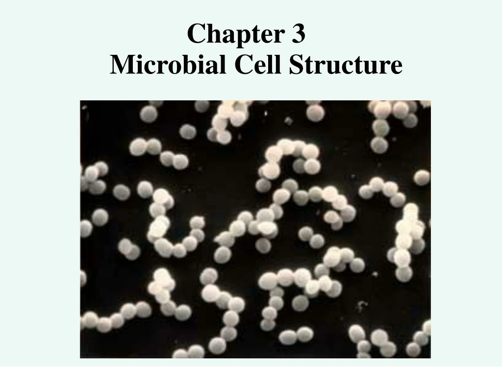
Examples of Capsule Forming Bacteria Streptococcus pneumonia: A polysaccharide capsule that is poorly antigenic protects the bacterium from the host immune system and makes it more virulent. Streptococcus mutans: A dextran capsule (polysaccharide) helps the bacterium to stick to the surface of a tooth, calcium leeched from the enamel mineralizes the capsular matrix. Xanthomonas campestris: A soil bacterium that causes soft rot of vegetables is protected from freezing and desiccation (drying) by a polysaccharide capsule. Streptococcus cells negatively stained to show capsules
Bacterial Capsules Which ONE of the following statements about bacterial capsules is FALSE? A. Most bacterial capsules are made of DNA. B. A capsule may help a bacterial cell to survive exposure to temperatures below 0oC. C. A capsule may help a bacterial cell stick to surfaces or other bacterial cells. D. A capsule may help to protect a bacterial cell from drying out. E. A capsule may help a pathogenic bacterial cell to evade host phagocytic white blood cells.
Bacterial Capsules Which ONE of the following statements about bacterial capsules is FALSE? A. Bacterial capsules are made of DNA. FALSE Most bacterial capsules are polysaccharide, butsome are amino acid polymers, others hydrocarbon. B. A capsule may help a bacterial cell to survive exposure to temperatures below 0oC. C. A capsule may help a bacterial cell stick to surfaces or other bacterial cells. D. A capsule may help to protect a bacterial cell from drying out. E. A capsule may help a pathogenic bacterial cell to evade host phagocytic white blood cells.
Bacterial Appendages Structures that are found on the outside of the cell Bacterial appendages are simple protein tubules. - Flagella: rotating helical fibers that act as propellers Monotrichous (one hair) Lophotrichous (a tuft or clump of hair) Amphitrichous (on both sides) Peritrichous (all around) Axial Filaments (inside the periplasmic space) - Fimbrae (or Pili): fibers that help bacteria stick to surfaces and to each other - Sex Pilus: a special fimbri that is used during conjugation
Bacterial Flagella A bacterial flagellum consists of a single protein fiber that is external to the cell membrane, it is a helical tube of protein that rotates like a propeller or corkscrew.
Axial Filaments are specialized flagella seen within the periplasmic space (endoflagella) of Spirochetes. (The Spirochetes includes such human pathogens asTreponema palidium, the causative agent of syphilis &Borrelia burgdorferi, causative agent of Lyme disease.) Image from:classes.midlandstech.edu
Bacterial Appendages - Continued Pili or Fimbrae - Pili are hollow tubes of protein that stick out on the outer surface of bacteria. - Pili are about the same diameter as flagella but are shorter and straight. - Pili help bacteria to stick to surfaces and to each other. - Some pili are virulence factors that allow pathogenic bacteria to stick to host cells. - Other pili are important for the formation of biofilms. - In some strains of E. coli there is a special pilus made called the F-pilus. This pilus causes two cells to stick together so DNA can be transferred from one to the other by conjugation. (F is "fertility".)
Bacterial Cell With Fimbrae Fimbrae help some E.colistrains to stick to cells that line the urethra.This is an important factor in urinary tract infections. diagram from Urology 7th ed. by Campbell
Bacterial Cell PhysiologyThe Envelope: cytoplasmic membrane always present cell wall usually present (peptidoglycan) capsule sometimes present appendages sometimes present flagella, pili, fimbraeThe Cytoplasm: nucleoid always present ribosomes always present plasmids often present endospore sometimes present chromatophore sometimes present inclusion bodies sometimes present starch granules, polyphosphate granules iron granules, sulfur granules, storage proteins
Structures of the Bacterial Cytoplasm Nucleoid: a region of cytoplasm where the DNA is located, it is not surrounded by a membrane
Ribosomes Enzyme complexes that read mRNA and, with the tRNAs, make proteins.
Cytoplasmic Structures: Bacterial Endospore The endospore is a tough resting state that allows bacteria to survive a period of harsh conditions such as when the soil dries out or they run out of food. The endospore is not a reproductive structure because sporulation begins with a single vegetative cell and ends with a single spore. Endospores are the toughest form of life known on Earth. Spores are very resistant to: heat, radiation, dessication, lack of nutrients, antimicrobial chemicals and physical abrasion. Spores can survive in an inert state for many years, even centuries. Endospore Structure The endospore coat is a highly modified cell wall that contains special coat proteins and dipicolinic acid in addition to peptidoglycan. Dipicolinic acid chelates calcium ions, so the spore coat is somewhat mineralized. The core of the spore contains everything found in the cytoplasm of a vegetative cell except it is very nearly metabolically inert and it contains much less water than normal cytoplasm.
Most spore forming bacteria belong to one of only two genera: Bacillus and Clostridium. Both are Gram + rods. Bacillus grows aerobically, while Clostridium is anaerobic. Anthrax is caused by inhalation of spores from Bacillus anthracis. Tetanus and gas gangrene are caused by contamination of a wound site by Clostridium spores. Botulism is caused by eating food that is contaminated by a toxin that is made by Clostridium botulinum. You can find more about endospores in your textbook on pages 316 – 319 including figures 11.8 and 11.9.
Cytoplasmic structures: The Chromatophore The chromatophore is a membrane structure found only in photosynthetic bacteria that contains light harvesting pigments. The pigments allow the cell to absorb sunlight and produce energy. Chromatophore means "colored body", from the color of the light harvesting pigments (green, purple, yellow-green).
Photosynthetic bacteria produce light harvesting pigments called bacteriochlorophyll and carotenoids. Bacteriochlorophylls are similar to plant chlorophyll except they often absorb different wavelengths of light. This means that photosynthetic bacteria are different colors. Many photosynthetic bacteria are purple or yellowish green rather than the more bluish-green of land plants. Also unlike plants, bacterial photosynthesis does not always involve the production of oxygen.
Inclusion bodies are insoluble particles that accumulate in some bacterial cells under certain conditions including: media recipe, availability of specific nutrients or oxygen and culture age. Some inclusion bodies can be visualized using special stains. Inclusion bodies are usually some form of stored material. Some inclusion bodies are metabolic waste products while others are stored nutrients. Some bacteria accumulate starch granules, others accumulate granules of sulfur, polyphosphates or poly-hydroxybutyrate.
Common Types of Inclusion Bodies (Granules) That Are Found in Bacterial Cells 1. Starch (a glucose polymer)2. Protein (storage)3. Lipoid granules (globs of fat)4. polyhydroxybutyrate5. sulfur6. iron
Differences Between the Archaea and the Bacteria 1. Cell Membrane Lipids – Fatty acids in bacterial lipids have straight chains, fatty acids in Archaea have branching chains. 2. Cell Wall Composition – Bacterial cell wall is made of peptidoglycan, Archaeal cell wall is made of other compounds. (pseudopeptidoglycan – lacks n-acetyl muramic acid) 3. RNA polymerase in Archaea is more like eukaryotic cells. DNA polymerase in Archaea is more like eukaryotic cells 4. Currently, there are no Archaea that are known to cause human disease, but there are more than 300 species of bacteria that are known to cause disease in humans
Archaea Which ONE of the following statements about Archaea is TRUE? A. Archaeal cells have well-defined nuclei that are surrounded by a nuclear membrane. B. All known Archaea species are human pathogens. C. The phospholipids found in Archaeal cell membranes are exactly like those of bacterial cell membranes. D. The ribosomes from Archaea are about the same size as those from bacterial cells.
Archaea Which ONE of the following statements about Archaea is TRUE? A. Archaeal cells have well-defined nuclei that are surrounded by a nuclear membrane. B. All known Archaea species are human pathogens. C. The phospholipids found in Archaeal cell membranes are exactly like those of bacterial cell membranes. D. The ribosomes from Archaea are about the same size as those from bacterial cells.
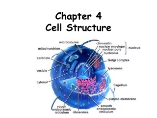
Structures Seen in Eukaryotic Cells But Not in Prokaryotes - Endoplasmic Reticulum - Cytoskeleton - Mitochondria - Phagolysosomes - Chloroplasts - snRNPs - Nuclear Membrane The Endoplasmic Reticulum Endo- : inside -plasmic: the stuff, matter Reticulum: net The E.R. is a complex network of internal cell membranes that is attached to both the cytoplasmic membrane and the nuclear membrane. Ribosomes are often attached to the endoplasmic reticulum.
Mitochondria Mitochondria are membrane bound "energy producing factories" that are not seen in bacteria. Most mitochondria are rod shaped and they are the size of small to average bacterial cells. Mitochondria are essential for survival of most types of eukaryotic organism (there are a few strange protozoa that lack them). Mitochondria have a double membrane.The enzymes of the electron transport chain are found in the inner mitochondrial membrane.Mitochondria consume oxygen.