Download
1 / 5
50 likes | 197 Views
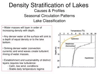
This guide outlines a method for determining the tissue volume density of human testes by counting specific structures through image analysis techniques. Using a Plus Counter transparency sheet, magnify online images of the human testes and precisely count the number of structures, including seminiferous tubules, Leydig cells, and blood vessels. Record your data, repeat counts in different areas, and calculate the tissue volume density based on your hits. This systematic approach facilitates a clearer understanding of testicular composition and its cellular distribution.

E N D
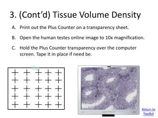
3. (Cont’d) Tissue Volume Density Print out the Plus Counter on a transparency sheet. Open the human testes online image to 10x magnification. Hold the Plus Counter transparency over the computer screen. Tape it in place if need be. Return to Toolkit
3. Tissue Volume Density Print out the Plus Counter on a transparency sheet. Set the magnification to 80x. Hold the Plus Counter transparency over the computer screen. Tape it in place if need be. Count the number of structures under the center of each cross (one “hit” is scored for each cross). Structure will be either: Seminiferous Tubules, Boundary Tissue of Seminiferous Tubules, Leydig Cells, or Blood Vessels. Record the numbers in the worksheet and add up totals. Move to a new area and repeat hit count 4 or more times. Add up the total hits that land on each structure from all repeated counts, divide by the number of possible “hits” (e.g., 5 repeats X 25 pluses per sheet = 125), and multiply that value by 100 to calculate tissue volume density.
Human Testes (number of hits on structures) 21 Seminiferous Tubules, 2 Boundary Tissue, 2 Leydig Cells, and 1 Blood Vessel Human Testes Image
3. Tissue Volume Density Example Data Leydig Cells Seminiferous Tubules Blood Vessels Boundary Tissue 21 21 20 21 21 3 3 2 3 3 3 2 3 2 3 0 0 0 1 0 Total Hits 104 14 1 13
3. Tissue Volume Density Example Data:percentage of whole occupied by each component Blood Vessels Seminiferous Tubules Boundary Tissue Leydig Cells Total Hits Possible Hits 5 counts x 25 pluses per sheet = 125 Volume Density of Tissue 13 1 14 104 Conclusion: The bulk of volume of testicular tissue is occupied by seminiferous tubules, Leydig cells and tubular boundary tissue contribute about 10% each, and a low volume of blood vessels. 104 14 13 1 x 100 = x 100 = x 100 = x 100 = 125 125 125 125 11% 83% 0.008% 10%