Dual-Force Convolutional Neural Networks for Accurate Brain Tumor Segmentation
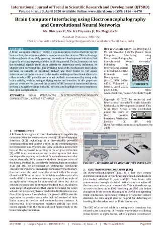
This paper presents a novel dual-force convolutional neural network approach for accurate brain tumor segmentation, leveraging multi-level information from MRI scans. The study addresses the limitations of existing methods like DeepMedic and U-Net by introducing a dual-force training scheme and a label distribution-based auxiliary loss function. Experiments on BRATS 2015 and BRATS 2017 datasets demonstrate significant improvements in segmentation accuracy. This method enhances the quality of features learned from deep models, promoting effective differentiation between tumor and normal tissues.

Dual-Force Convolutional Neural Networks for Accurate Brain Tumor Segmentation
E N D
Presentation Transcript
Dual-force convolutional neural networks for accurate brain tumor segmentation Shengcong Chen, Changxing Ding, Minfeng Liu 2018
Introduction • Median Survival Time of High-Grade Gliomas patients is less than 2 years • Median Survival Time of glioblastoma patients is 4.9 months • Magnetic Resonance Imaging (MRI) is widely used for diagnosis and treatment of brain tumors • MRIs are large-volume 3D scans • manual segmentation is time-consuming and tedious • manual segmentation is likely to be affected by raters’ personal experience • designing an accurate brain tumor segmentation system is a challenging problem • shape and internal structures of tumors are varied and complicated • surrounding normal tissues are also in a wide variety of appearance due to variable locations of tumors and the so-called tumor mass effect • boundaries between normal tissues and tumor tissues tend to be obscure and therefore they are difficult to differentiate • State-of-the-art dense prediction networks include DeepMedicand U-Net
Introduction • DeepMedic • utilizes feature maps extracted from the final convolutional layer to predict the labels of voxels within the central regions of input patches efficiently • final convolutional layer in CNN usually focuses on extracting high-level semantic information. The lack of low- and middle-level information restricts the performance of DeepMedic • U-Net • They consist of an encoding network with down-sampling operations and a de-coding network with up-sampling or deconvolutional operations • U-Nets introduce feature maps in the encoding network to the de-coding network through successive concatenation operations in order to introduce multi-level information gradually and finally promote the prediction accuracy
Introduction • a series of approaches to enhance the quality of the learnt features • extend the popular DeepMedic model to Multi-Level DeepMedic to make use of multi-level information for more accurate segmentation • a novel dual-force training scheme to promote the quality of multi-level features learnt from deep models. It is a general training scheme and can be applied to many exiting architectures, e.g., DeepMedic and U-Net • design a label distribution-based loss function as an auxiliary classifier to encourage the high-level layers of deep models to learn more abstract information • a novel Multi-Layer Perceptron-based post-processing approach to refine the prediction results of deep models
DeepMedic • For simplicity, only the full-resolution pathway of DeepMedic is shown. Each Conv block in the figure refers to the combination of one convolutional layer, one batch normalization layer and one ReLU layer.
Multi-level DeepMedic Feature map patches are cropped from five selected layers and then concatenated to compose multi-level features. several crop layers to extract multi-level features of the same size, i.e., 9 ×9 ×9 from five selected layers of DeepMedic. Each crop layer crops a 3D patch from the center region of the feature map. To fuse these multi-level information, the cropped feature map patches are first concatenated and then fed into three successive 1 ×1 ×1 convolutional layers for final prediction.
U-Net • U-Net comprises an encoding network and a decoding network. • In encoding network, stacked convolutional layers are followed by pooling layers to compress the size of feature maps and extract high-level semantic information. • The decoding network incorporates a set of deconvolutionallayers for resolution recovery. Moreover, feature maps generated by deconvolutional layers are concatenated with those produced by convolutional layers of the encoding network with the same resolution. The combination of feature maps enables U-Net to gradually fuse multi-level information for accurate brain tumor segmentation.
U-Net • Important model parameters of 3D U-Net adopted in this paper. Filter number in the last convolu- tional layer depends on the number of normal and tumor classes, i.e., 4 (BRATS 2017) or 5 (BRATS 2015) in this paper.
DF-MLDeepMedic • The auxiliary loss function is attached to the ‘Conv8’ layer of the full-resolution pathway. For the two-pathway DF-MLDeepMedic, there is another auxiliary loss function attached to the deconvolutional layer of the low-resolution pathway.
DF-U-Net • A new convolutional classification layer and the auxiliary loss function is attached to the ‘Conv8’ layer.
Experiments • Data: • two brain tumor segmentation datasets: • BRATS 2015 • 274 3D MRI scans • 4 classes of tumor tissues: • necrotic, edema, non-enhancing tumor, and enhancing tumor • BRATS 2017 • 285 3D MRI scans • 3 classes of tumor tissues: • necrotic, edema, and enhancing tumor • Image Size: 240 × 240 × 155 • Train: 255 images • Validation: 30 images
Experiments • Implementation details • open-source deep learning package C3D • Batch size: 64 • Learning rate: 0.001 • maximum number of iterations: 40 epochs
Experiments • Evaluationcriteria • Performance comparison between DeepMedic and MLDeepMedic on the local validation subset of BRATS 2017 (%)
Experiments • Performance comparison between DeepMedic, MLDeepMedic, and DF-MLDeepMedic on the local validation subet of BRATS 2017 (%).
Experiments • Performance comparison between U-Net and DF-U-Net on the local validation subset of BRATS 2017 (%). • Performance Comparison of Different Post-Processing Methods on Local Validation Dataset of BRATS 2017 (%).
Experiments • Comparison of Dice scores between DFNs and the state-of-the-art methods on BRATS 2017 Online Validation Dataset (%). • Comparison of Dice scores between DFNs and the state-of-the-art methods on the BRATS 2015 online test dataset (%).
Experiments • Columns: • original MRI slices • ground-truth segmentation • DeepMedic • MLDeepMedic • DF-MLDeepMedic (S) • DF-MLDeepMedic (L) • Colors: • Edema (green) • enhancing tumor (red) • necrosis and non-enhancing tumor (blue) 1 2 3 4 5 6
Conclusion • Accurate brain tumor segmentation depends on multi-level information • a dual-force training strategy is proposed to explicitly encourage deep models to learn high-quality multi-level features by a label distribution-based loss function to learn the abstract semantic information and a softmax loss function for segmentation using multi-level features • Applying the proposed strategy to deep models only slightly increases the time and space complexity while training • an MLP-based post-processing method is also proposed that can automatically learn post-processing rules from data rather than manual summarization • One shortage of the MLP-based post processing method is that its training process is separated from that of DFN; therefore, the entire framework is not completely end-to-end