Download
1 / 31
310 likes | 331 Views
Explore a detailed case study involving a patient with ataxia, genetic testing, autopsy findings, and a diagnosis of Niemann-Pick disease type C1. Uncover the complexities of this rare condition through systematic genetic analysis.

E N D
Diagnostic slide session 2010American Association of Neuropathologists Case 2010-12
ContributorsArnulf H. Koeppen, Ashley N. Davis, Jennifer A. Morral, Edward S. Johnson, Jiang Qian, Kinuko Suzuki, Uta Gölnitz, Matthias Wittstock, Arndt Rolfs, and Richard Camicioli
Clinical History Main problem: Ataxia beginning at age 10 Family history: Tremor in mother and maternal grandfather Genetic testing: Normal for SCA 1, SCA-2, SCA-3, SCA-6, and Friedreich’s ataxia Neurological findings: Normal mental status; saccadic intrusions into ocular pursuit movements; ataxia; dysmetria; dysarthria; hearing loss; modest hyperreflexia; and a right Babinski sign. Course: Relentless progression to intense rigidity of her extremities; dystonia; leg spasticity; and sustained ankle clonus; death at 39 Imaging: Magnetic resonance imaging unrevealing. Autopsy findings: Pulmonary congestion and an angiomyolipoma of the right kidney; brain weight 1321 g; substantia nigra pale.
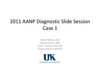
Oculomotor nucleus; PAS Oculomotor nucleus; HE 100 μm
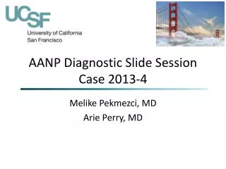
Thalamus Parahippocampal gyrus Globus pallidus; PAS
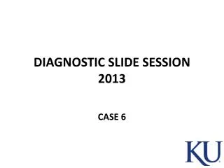
LAH Nucleus of Onuf
This case GM2 gangliosidosis :Sandhoff’s d.
Systematic genetic analysis of possible GM2 gangliosidosis (Institute of Molecular Diagnostics, Rostock, Germany)Patient’s DNA:(1) Hex A: normal, excluding Tay-Sachs disease(2) Hex B: normal, excluding Sandhoff’s disease(3) GM2A: normal, excluding Tay-Sachs variant
Systematic analysis of Niemann-Pick type C1 disease (NPC1)Father’s DNA: R935Q ( known pathogenic mutation)Mother’s DNA: G992R (known pathogenic mutation)Patient’s DNA: R934Q/G992R (compound heterozygote of two known pathogenic mutations)
Genetic diagnosis:Niemann-Pick disease, type C1, OMIM 257.220Unusual: compound heterozygosity
20 μm Cortex; filipin
Acknowledgment. The neuropathological work was completed in the laboratories of VA Medical Center in Albany, N.Y. (AHK); Albany Medical College (JQ); and WC Mackenzie Health Sciences Center, Edmonton, AB, Canada (ESJ). The mutations were identified at Centogene and University of Rostock, Rostock, Germany (UG, MW, AR). RC contributed the clinical data.