Download
1 / 16
170 likes | 356 Views
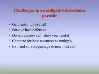
Obligate Intracellular Organisms. Bacterial Intracellular Organisms. Bartonella spp. Pathogenesis not well understood. Adheres to endothelial cells and is engulfed. Chlamydiae spp M. Tuberculosis Legionella spp Toxoplasma gondii. Lives in a phagosome & prevents phagolysosomal

E N D
Bacterial Intracellular Organisms • Bartonella • spp Pathogenesis not well understood. Adheres to endothelial cells and is engulfed • Chlamydiae spp • M. Tuberculosis • Legionella spp • Toxoplasma gondii Lives in a phagosome & prevents phagolysosomal fusion Intracellular organism Escapes from phagosome & lives in cytosol • Ricketssiae • Listeria • shigella Lives in phagolysosome • Leishmenia • Coxiella
Chlamydia Species • Obligate intracellular organisms • Small round to ovoid cells, 0.3µm • Cell has peptidoglycan and an outer lipid layerresembling that of a Gram negative bacteria • Genome much smaller than that of other bacteria • They cannot make ATP (adinosine triphosphate) – dependent on host cell for energy production • They import nutrients from the cytosol into the endosome with the help of tubular projections on the surface of the reticulate body
Chlamydia Species • Growth characteristics: cannot be grown on artificial bacteriologic medium, only grows within living tissue or tissue culture cells • Replication is by binary fission but they undergo morphologic variation during replication cycle • have distinct elementary bodies (EB) adapted for extracellular survival and initiation of infection and • reproductive reticulate body (RB) forms for intracellular replication
Chlamydia Species • Antigenic composition: • Serotyping is based on specific cell wall proteins • Each serotype is associated with a specific disease
Epidemiology • Chlamydiae are susceptible to environmental conditions, survive only a short time outside the host • Transmission is by direct contact among humans • C. psittaci is pathogenic for birds and domestic fowl and is transmitted to humans by inhalation of bacteria in droplets or dust
Pathogenicity • Highly infectious • Pathogenesis of disease caused by these bacteria is not well understood • Different strains of both C. trachomatis and C. psittaci show different degrees of virulence • Phagocytosed chlamydiae prevent fusion of lysosome to the phagosome thus escape intracellular destruction by lysosomal enzymes • Produce heat-labile toxin • Competition for nutrients • C. trachomatis may exist in a latent state and may reactivate if host becomes immunosuppressed
Diseases caused by Chlamydia species • Chlamydia cause persistent and recurrent infections • Three species cause human disease: • C. trachomatis: trachoma a chronic infection which causes blindness, inclusion conjunctivitis an acute infection, urethritis, cervicitis, salpingitis and lymphogranuloma venerium (LGV) • C psittaci: which causes pneumonia, • C. pneumoniae: which causes pneumonia, pharyngitis, bronchitis
Laboratory Diagnosis and Treatment • C. trachomatis is detected using • Direct fluorescent antibody staining of genital exudates with fluorescein-labelled monoclonal antibodies against MOMP or LPS. • Serologic techniques are not used for C. trachomatis and culture is rarely done. • Nucleic acid hybridization techniques are widely used to detect asymptomatic genital infection in women. • C. psittaci pneumonia: a 4 fold rise in CF antibody to chlamydial group antigen • Treatment of Chlamydia includes macrolides and tetracyclines
Obligate intracellular pathogens Small GNB 0.3-0.5µm Stain poorly with bacteriologic stains Visualized easily with Giemsa Important members include: R. prowazekii, R. typhi, and R rickettsii Rickettsia
Growth characteristics • Only grows within living tissue or tissue culture cells • Intracellular growth: enter host by endocytosis, produces a phospholipase which destroys the phagosome and it begins to grow in the cytoplasm of host cell. • require coenzyme A, nicotinamide-adenine dinucleotide (NAD), + energy from host cell • Free rickettsiae: cease metabolic activity and begin to leak intracellular constituents resulting in lack of infectivity within a short period
Generally have animal reservoirs Transmitted by the bite of an arthropod vector Pathogenicity
Bacteria invade the vascular epithelial cells and become widely disseminated Damage to the endothelial cells in small vessels causes vasculitis, obstruction, capillary leaks and thrombosis. This causes a rash, organ damage and potentially shock. Pathogenesis
Spotted fever Fever, headache and rash Eschar = black scar at site of bite Clinical Disease
Diagnosis and Treatment • By necessity, the diagnosis must be made clinically • If a rickettsial illness is suspected, treat first with an antibiotic that can reach a therapeutic concentration inside infected cells (tetracyclines, long acting macrolides, fluoroquinolones etc.), and then confirm the diagnosis with serology.