Download
1 / 51
530 likes | 631 Views
Exocytosis, endocytosis, vesicular transport. Anna L. Kiss Deparment of Anatomy, Histology and Embryology, Semmelweis University Budapest 2017. Endocitózis / exocitózis. Endosome. Endosome : Content: gradually acidifying proton pump in their membrane.

E N D
Exocytosis, endocytosis, vesicular transport Anna L. Kiss Deparment of Anatomy, Histology and Embryology, Semmelweis University Budapest 2017
Endosome Endosome: Content: gradually acidifying proton pump in their membrane. Acidic pH: receptor-ligand disszociation futher fate can be separated
Endosome Endosome: cellular organelles with variable size and shape – close to the plasme membrane. early or sorting endosome recycling endosome Multivesicular body /vs late endosome lysosome
Lysosomes EM: membrane surruonded vacuoles, fine granular structure inside. Content: acidic hydrolytic enzymes (more than 40), pH optimum: pH 5. Function: degradation of macromolecules by hydrolysis.
Lysosomes I. Primary lysosomes:contain ONLY lysosomal enzymes Lysosomal enzymes: nucleases, proteases, glycosidases, lipases, phosphatases, sulphatases, …(more than 40). One lysosome can contain more enzymes! Acidic pH : H+ pump (proton- ATPase, vacuolar type) in the membrane of the lysosomes. Lysosomal membrane (and the neutral pH of the cytosol) protect the cell from the self-digestion. Membrane protection: highlyglycosylated proteins and lipids in the membrane ((sugar chains prevent direct contact of the enzymes with the membrane). Transport proteins and proton-ATPase in the membrane. II. Secondary lysosomes
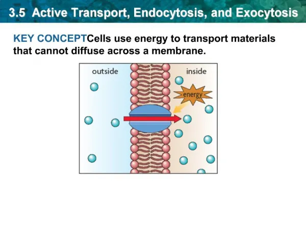
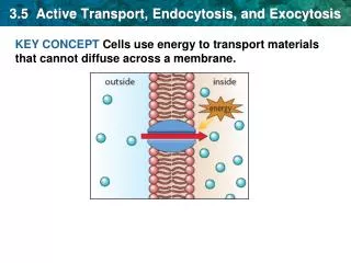
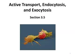
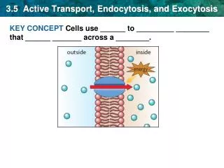
Exocytosis: transport from the cytoplasm to the cell membrane Sorting from the Golgi network • To the plasma membrane A. Membrane proteinsreleased to the plasma membrane from the trans-Golgi network by vesicles, continuous, vesicles open by fusion B. Secretory proteins containing vesicles, vacuoles pinch off from the trans-Golgi network, open by membrane fusion, their contant is released Regulated exocytosis: exocytosis stimulated by signal (signal transduction) mast cell constitutive exocytosis: continuous, without signal cell membrane secretion (regulated exocytosis) constitutive exocytosis trans-Golgi network lysosome 2. To the lysosome (or late endosome) Lysosomal proteins: localization signal: mannose-6-phosphate, receptors: man-6-P-receptors rER nuclear membrane
Exocytosis Membrane fusion: phospholipids of the membranes fuse with each other
On the way from Golgi to plasma membrane along microtubulus
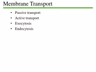
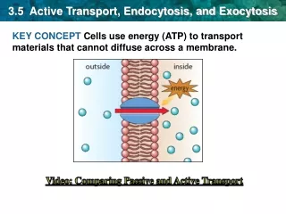
Endocytosis • Endocytosis: • - activetransport • - uptake of substancesintothecellbyinvaginationoftheplasmamembrane • - the endocytosed substanceorparticle is separatedfromthecytosolby a membrane (derivative of theplasmamembrane). • Phagocytosis • Pinocytosis
Phagocytosis Endocytosis of large particles (>0,1-0,5 μm) Examples:macrophages and neutrophilic granulocytes (professional phagocytes) bacterium cytoplasmic process macrophage plasma membrane erythrocyte Phagocytosis of bacteria into a blood leukocyte (neutrophilic granulocyte). Electron micrograph. A macrophage engulfs two erythrocytes. Arrow: margin of the cytoplasmic process. Scanning EM.
Phagocyted particles: debris of dead cells, bacteria, senescent erythrocytes, apoptotic cells and bodies, foreign particles, etc. … Fc-ends of IgG molecules Phases of phagocytosis: Fc-receptor bacterium 1. Adsorption: particles to be phagocytosed are bound to the cell surface (mostly to receptors in the plasma membrane). Specific receptors: e.g. Fcγ-receptors, complement-receptors. Example: phagocytosis of bacteria covered with immunoglobulin molecules (IgG) into a macrophage cell. Non-specific receptors:(scavanger receptors): receptors recognizing unusual carbohydrates or a changed phospholipid pattern on the surface Living cells are not phagocytosed due to proteins on their cell surface which activate inhibiting receptors on the plasma membrane of the phagocytic cell (inhibition of phagocytosis). plasma membrane actin microfilaments 2. Ingestion: the phagocytic cell with its collar-like cell process (pseudopodium, lamellipodium) surrounds the particle, the free margins of the process are closed over the particle. Actin microfilaments are polymerised in the cell process. After complete closure, the particle is enclosed into a vacuole which becomes detached from the plasma membrane. Phagosome.
II. Pinocytosis Uptake of particles smaller than 0,1 μm, or macromolecules or dissolved substances by endocytosis. The resulting vesicles are 50-100 μm large. • Fluid phase pinocytosis: the particles are in a dissolved state (solution) and are taken up into the cell by invagination of the plasma membrane (endocytic vesicles). • Adsorptive pinocytosis. The small particles are bound to the plasma membrane (enrichment of the particles on the surface of the plasma membrane), therefore uptake is more effective even at low concentrations of the particles. • Non-specific binding sites (e.g. glycocalyx, non-specific receptors). Some bacterial toxins (Diphteria), viruses (e.g. Influenza, VSV, Semliki Forest virus) are taken up into the cell this way. • Specific binding sites (receptors): receptor-mediated pinocytosis (or endocytosis). Ligands: immunoglobulins, α2-makroglobulin, transferrin, LDL , asialoglikoproteins, … Receptors: integral membrane proteins
Endocytoticpathways Phagocytosis(„eating”): >0,25µm Pinocytosis („drinking”) Macropinocytosis: >1µm Endocytosisviaclathrin-coatedvesicles: ~120nm Endocytosisvia caveolae: ~60nm Endocytosis clathrin- és caveolin-independent and/ordiynaminindependent: ~90nm Regulated portals of entry into the cell SEAN D. CONNER AND SANDRA L. SCHMID Nature 422, 37 - 44 (06 March 2003)
Macropinocytosis Depends on ruffling
Endocytoticpathways Phagocytosis(„eating”): >0,25µm Pinocytosis(„drinking”) Macropinocytosis: >1µm Endocytosisviaclathrin-coatedvesices: ~120nm Endocytosisvia caveolae: ~60nm Endocytosis clathrin- és caveolin-independent and/ordiynaminindependent: ~90nm Regulated portals of entry into the cell SEAN D. CONNER AND SANDRA L. SCHMID Nature 422, 37 - 44 (06 March 2003)
Receptor-mediated endocytosis via clathrin coated vesicles Highly regulated, specific uptake precess! Clatrhin-coated vesicles: a basket-like coat is forming around the vesicle Clathrin: heavy and light chain triskelion pentagons and hexagons
Clatrhincoatedvesiclesmediatedendocytosis coated pit Receptor proteins with the bound substance, adaptin proteins and clathrin assemble in the membrane. The GTP-binding protein dynamin separates the vesicle from the plasma membrane. The clathrin coat is then shed from the vesicle in the cytosol.
Clathrin-mediatedendocytosis Pinching off the vesicle by dynamin (small GTP-ase)
Clathrin-coated pits and vesicles under the cell membrane, seen from inside the cell, electron micrograph, freeze fractured and deep etched Heuser technique. clathrin-coated vesicle clathrin-coated pit)
Endocytoticpathways Phagocytosis(„eating”): >0,25µm Pinocytosis („drinking”) Macropinocytosis: >1µm Endocytosisviaclathrin-coatedvesices: ~120nm Endocytosisvia caveolae: ~60nm Endocytosis clathrin- és caveolin-independent and/ordiynaminindependent: ~90nm Regulated portals of entry into the cell SEAN D. CONNER AND SANDRA L. SCHMID Nature 422, 37 - 44 (06 March 2003)
Caveolae-mediatedendocytosis Caveolae: d: 50-100nm flask-or omega-shaped invaginations Dr L. Kiss Anna felvétele Dr L. Kiss Anna felvétele
Caveolae: caveolin-containinglipidrafts • Highlyhidrophobicmembranedomenes: cholesterol, sphyngolipids, glycosyl-phosphatydil-inositol; lipidrafts • Caveolin: • caveolin-1 • caveolin-2 • caveolin-3 • Cavin (PTRF): accessory protein
Caveolae • Lipidrafts: highlyhydriphobicmembranedomains • richinsphyndolipids, cholesterol, glycolipids • proteins: caveolin (1, 2, 3) Lipidraft is invaginating
Caveolae-mediatedendocytosis • Uptake of SV40 virus : occurswith caveolae • Virusbindsto MHC I presentinthehostcellplasmamembrane • MHCI (the receptor forvirus) is presentin caveolae • Caveolin is highlyphosphorylatedbySrckinase, (tyrosinephosphorylation) • Reorganization of thecorticalcytoskeleton (actinfilaments) actin-tail is formed • Dynamin(smallGTPase) associatestotheneckregion of caveolae • Caveola is pinchingoff
Caveola: „signallingorganelle” scaffolding domene: at the N terminus; binds signaling molecules
Endocytoticpathways Phagocytosis(„eating”): >0,25µm Pinocytosis („drinking”) Macropinocytosis: >1µm Endocytosisviaclathrin-coatedvesices: ~120nm Endocytosisvia caveolae: ~60nm Endocytosis clathrin- és caveolin-independent and/ordiynaminindependent: ~90nm Regulated portals of entry into the cell SEAN D. CONNER AND SANDRA L. SCHMID Nature 422, 37 - 44 (06 March 2003)
The fate of pinocytotic vesicles and of the interiorized substance. The pinocytotic vesicle sheds its coat and fusions with the early endosome. Endosome: A membrane-bound cell organelle of variable size and shape. Its interior is gradually acidified by the action of proton pumps in the endosomal membrane. Early endosomes: mostly present in the marginal cytoplasm. pH-values 6.5-5, at this pH most ligands are released from their receptors (ligand-receptor uncoupling). Important sorting organelle towards plasma membrane, (recycling endosome) Golgi-apparatus and late endosome – lysosome. Late endosomes: more deeply situated in the cytoplasm and having an acidic pH. Lysosomal enzymes begin to accumulate by a vesicular transport from the trans-Golgi network. Multivesicular bodies: the membrane of late endosomes is budding into the interior of the organelle to form vesicles (many vesicles in the lumen). This way integral membrane proteins can be degraded by lysosomal enzymes in the organelle. Lysosomes: mostly close to the Golgi-apparatus, surrounded by a membrane and having a pH of 4,5-5. Many hydrolytic enzymes destined for degradation of organic substances.
Fate of the receptors • Recycling to the plasma membrane by a vesicular transport after releasing their ligands in the early endosome. Vesicles with empty receptors are budding off from the membrane of early (and partly also from late) endosomes and travel to the plasma membrane with which they fuse by exocytosis (receptor recycling). This way the membrane of vesicles (together with the receptors) is reintegrated into the plasma membrane. Example: LDL receptors. • They reach the late endosome and are degraded in lysosomes (receptor downregulation). Example: EGF receptor (epithelial growth factor with its receptor). Fate of the ligands • Degradation.The ligand is transported further from early endosome towards late endosome and lysosome where it is degraded. Or: • Recirculation to the cell surface. In early endosome the ligand remains bound to its receptor and recycles back to the plasma membrane together with its receptor. Example: transferrin (an iron-transporting protein which after releasing iron in the endosome recirculates together with its receptor to the plasma membrane ready for a further cycle).
Lysosomes • Primary lysosomes • Secondary lysosomes: primary lysosome+ phagocytotic vacuole heterophage vacuole autophage vacuole (phagocytosed particles: dead cells, bacteria aged red blood cells, apoptotic cells and bodies, foreign particles, etc.) autolysosome heterolysosome
Autophagy Focal digestion of cytoplasmic regions in unfavorable conditions („the cell eats itself”). Elimination of unnecessary and/or damaded cell components. - A C-shaped sequestration cisterna surrounds a cytoplasmic region to be degraded. Two parallel membranes, the inner membrane disappears later. - The cytoplasmic region, totally separated by the sequestration cisterna (autophagosome) is fused with a lysosome and degraded. A widespread autophagy can lead to death of the cell, many regard it as a second form of programmed cell death. Sequestration cisterna (arrows), electron micrograph. Autophagosome, electron micrograph Micrographs by Prof. J. Kovács
Transcytosis Transport of macromolecules in an intact form through the cell. Endocytosis → vesicular transport →early endosome→ vesicular transport→ exocytosis A special case of receptor-mediated endocytosis:the endocytosed molecules avoid lysosomal degradation.
Transcytosis Examples: • Uptake and transport of maternal IgG through the gut epithelium in newborns. • Uptake and transport of maternal IgG through the placental epithelium in the fetus. • Uptake and transport of IgA molecules through surface epithelia or in a secretory epithelium from the connective tissue space into the lumen. apical membrane domain gut lumen recycling transport vesicles early endosome Coated vesicles!! connective tissue space
Transcytosis Transport through capillary endothel Caveolae!!
Vesiculartransport 2 main moments: - pinching off the vesicles: signals, coat proteins - vesicular transport to the target membrane and fusion: regcognition molecules and fusion proteins
Vesiculartransport Formation of vesicles: coat proteins COPI, COPII: ER-Golgi clathrin: plasma membrane: vesicles transporting lysosomal emzymes caveolin signals: small G-proteins: GDP/GTP-binding proteins: Rab proteins: „identity” proteins ER-Golgi: ARP (donor membrane together with COP proteins) ARF: molekular switch (GTP-bound form: coat protein- membrane association GDP-bound form: uncoating, fusion can occur) Tethering and docking of the vesicles:fusion proteins: SNARE-s
Rab proteins kihorgonyozás dokkolás fúzió
Membranefusion trans-SNARE complex Fusion complex: excluding water membrane fusion