Medical Genetics
350 likes | 375 Views
Explore human karyotype, chromosomal banding, in situ hybridization, and chromosomal disorders. Learn about cytogenetics and molecular genetics in medical diagnostics and heredity.

Medical Genetics
E N D
Presentation Transcript
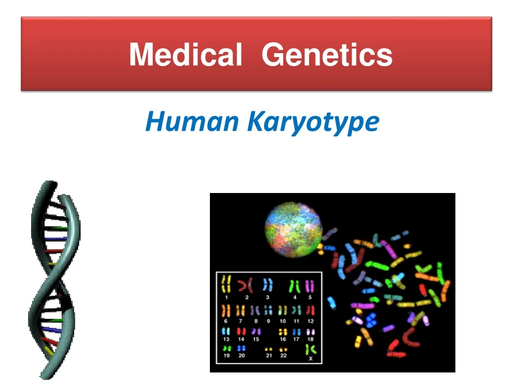
Medical Genetics Human Karyotype
Lecture Objectives: By the end of this lecture, the students should be able to: • Explain what a Karyotype is and how it is obtained. • Describe chromosomal banding and explain its use. • Describe the process of in situ hybridization and the information it provides.
GENETICS : ■ Cytogenetics: The study of the structure and function of chromosomes and chromosome behaviour during somatic and germline division ■ Molecular genetics: The study of the structure and function of genes at a molecular level and how the genes are transferred from generation to generation.
Cytogenetics: • Human Cytogeneticsinvolves the study of human chromosomes in health and disease. • Chromosome studies are an important laboratory diagnostic procedure in • prenatal diagnosis • certain patients with mental retardation and multiple birth defects • patients with abnormal sexual development • some cases of infertility or multiple miscarriages • in the study and treatment of patients with malignancies & hematologic disorders. • New techniques allow for increased resolution.
CHROMOSOMES: ■ carry genetic material ■ heredity: each pair of homologues consists of one paternal and one maternal chromosome ■ The intact set is passed to each daughter cell at every mitosis. EM of human chromosomes
The packaging of DNA:DNA coiling the visible structure of the chromosome Orders of DNA coiling and folding: • Primary coiling: DNA double helix • Secondary coiling: around histones (basic proteins) nucleosomes • Tertiary coiling chromatin fiber • Chromatin fibers form long loops on non-histone proteins tighter coils chromosome
Chromosomal disorders - These defects result from defects in the chromosomes. - Two groups: * Structural defects– defects in structure of chromosome. * Numerical defects– Increase or decrease in number of chromosomes - Chromosomal defects do not obey specific pattern of inheritance.
Chromosomal Disorders • The first chromosomal disorder was Trisomy 21 • (Downs syndrome) and was recognised in 1959. • These disorders are quite common and affect about • 1/800live born infants. • Account for almost half of all spontaneous • first-trimester abortions. • Do not follow a Pedigree pattern of inheritance.
KARYOTYPING • Defined as the mapping of CHROMOSOMES • 46 = (22x2) + X + Y • Conventional notation is “46,XY” or “46,XX” • G(iemsa)-banding, 500 bands per haploid recognizable • Short (“p”-etit) arm = p, other (long) arm = q The Giemsa stain, named after Gustav Giemsa, is a VERY common stain in pathology, often used to identify organisms in cells such as malaria and helicobacter, and MANY other things such as parts of cells and connective tissue. It is a VERY simple stain to do.
CLASSIFICATION OF KARYOTYPE • Cytogenetics: • ■ Non-Banded Karyotype • ■ Banded Karyotype • ■ High resolution Karyotype • Molecular cytogenetics: • ■ Fluorescent in situ hybridization (FISH).
Karyotype • A series of steps involved : • 1. Culturing • 2.Harvesting • 3.Slide-making • 4.Banding • 5.Staining • 6.Karyotyping • 7.Chromosome analysis
Procedure of Chromosome Preparation from Peripheral Blood 6 4 5 1.Culture media contains Phytohemagglutinin to stimulate T lymphocytes to divide 2 3 Prevents formation of the spindle arrest cell division during metaphase
B A C D E F G X Karyotyping • Based on: • the length • the position of the centromere • the presence or absence of satellites
Banding • Certain staining techniques cause the chromosomes to take on a banded appearance, • Each arm presenting a sequence of dark and light bands . • Patterns are specific and repeatable for each chromosome, • Allowing accurate identification and longitudinal mapping for locating gene positions and characterising structural changes. • Patterns, and the nomenclature for defining positional • mapping have been standardised
G Banding: Treat with trypsin and then with Geimsa Stain. R Banding: Heat and then treat with Geimsa Stain. Q Banding: Treat with Quinicrine dye giving rise to fluorescent bands. It requires an ultraviolet fluorescent microscope C Banding: Staining of the Centromere. Treat with acid followed by alkali prior to G banding R- banding and C-banding techniques are alternative methods that provide a reversal of the G- and Q-banding patterns. The ends of the chromosomes, which almost always stain positively, are well defined and thus terminal deletions are easily seen. .
Terminology. In Plate , a diagrammatic representation of chromosome 1 is labeled in the accepted format. Q bands are shown on the chromosome; however, G bands are identical except for the centromere. Each of the dark and light areas, or bands, is numbered, including the telomeres. The short arm of the chromosome is labeled "p," and the long arm "q," with the centromere in between the two. Each arm is divided into regions and numbered, with region I closest to the centromere. The bands within each region are also numbered, beginning with the band closest to the centromere. Sub-bands seen in high-resolution preparations are numbered in the same way and designated with a decimal point. Thus, 1p35 indicates the darkly stained band numbered 5 in region 3 on the short or p arm of chromosome 1. The notations 1p35.1 and 1p35.2 designate sub-bands within this band.
Banded Karyotype: Normal Banded Karyotypes A normal R-banded male Karyotype A normal G-banded male Karyotype
Fluorescence In-Situ Hybridization (FISH) Molecular cytogenetic techniques (e.g. FISH) are based on the ability of a single-stranded DNA probe to anneal with its complementary target sequence. They can be used to study chromosmes in metaphase or interphase. F.I.S.H. (gene “probes”) greatly enhances G-banding
Advances in the use of DNA probes have allowed cytogeneticists to hybridize these probes to chromosomes and determine if a specific DNA sequence is present on the target chromosome. This has been useful in detecting abnormalities beyond the resolution level of studying banded chromosomes at the microscope, and also in determining,the location of specificgenes on chromosomes.
Fluorescent In-Situ Hybridization • Uses fluorescent labelled DNA fragments, ~10,000 base pairs, to bind (or not bind) to its complement
FISH, used often in everyday pathology too, fluorescently “labels” pieces of DNA which connect to the corresponding strand during DNA replication. In situ hybridization (ISH) is a type of hybridization that uses a labeled complementary DNA or RNA strand (i.e., probe) to localize a specific DNA or RNA sequence in a portion or section of tissue (in situ) A DELETION in CHROMOSOME #22 TRIPLE CHROMOSOME #20
Fluorescence In-Situ Hybridization (FISH) FISH of interphase nuclei with a chromosome 21 centromeric probe showing 3 signals consistent with trisomy 21 FISH of metaphase with a probe for telomere showing signals at the end of each chromatid
FISH is POWERFULLY more sensitive, accurate, and specific, than G-banding. • SUBTLE MICRODELETIONS • COMPLEX TRANSLOCATIONS • AND TELOMERE ALTERATIONS
Common applications for FISH. Examples of diseases that are diagnosed using FISH include Prader-Willi syndrome, Angelman syndrome, 22q13 deletion syndrome, chronic myelogenous leukemia, acute lymphoblastic leukemia, Cri-du-chat, Velocardiofacial syndrome, and Down syndrome, but, IN GENERAL, diseases with partial or whole chromosome abnormalities.
Spectral karyotyping (SKY) is a laboratory technique that allows scientists to visualize all of the human chromosomes at one time by "painting" each pair of chromosomes in a different fluorescent color. Fluorescently labeled probes for each chromosome are made by labeling chromosome-specific DNA with different fluorophores.
SPECTRAL KARYOTYPING This technique is used to identify structural chromosome aberrations in cancer cells and other disease conditions when Giemsa banding or other techniques are not accurate enough. Each chromosome has a different color, sort of, although some of this is digital false color techniques, much in the same way, electron microscopy can generate “false” colors.
Items in the Description Of Karyotype: ■ Normal Karyotypes 46, XY 46, XX ■ Abnormal Karyotypes 47, XY, + 21 45, XO,- Y
Down Syndrome • 1 per 800 births • Large tongue • Flat face • Slanted eyes • Single crease across palm • Mental retardation • Some are not
Turner Syndrome • Short • Absence of a menstrual period • Produce little estrogen • Sterile • Extra skin on neck