Download
1 / 48
480 likes | 523 Views
Learn about foot & ankle exam, gait analysis, neurovascular assessment, landmarks for injections, common pathologies, and treatment options in this thorough guide.

E N D
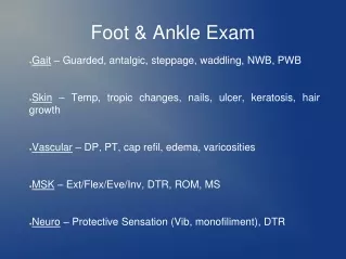
Foot & Ankle Exam Gait – Guarded, antalgic, steppage, waddling, NWB, PWB Skin – Temp, tropic changes, nails, ulcer, keratosis, hair growth Vascular – DP, PT, cap refil, edema, varicosities MSK – Ext/Flex/Eve/Inv, DTR, ROM, MS Neuro – Protective Sensation (Vib, monofiliment), DTR
Foot & Ankle Exam Normal Gait. Neurovascular status grossly intact. Protective Sensation intact. Dry Supple skin. There is no sign of infection. There is no edema. There is no erythema. There is no ecchymosis. There is no bony point tenderness. Rectus foot type. ROM of ankle is 20° df and 45° pf. STJ ROM is 30° inversion and 20° eversion. ROM of 1st MTPJ is 70° df and 25° pf. MS is 5/5. There is no proximal tenderness. The calf is supple and nontender. There is no anterior draw. Radiographs 3 views weightbearing of the ankle show no acute fracture or degenerative changes.
Foot & Ankle Landmarks Ankle Joint Injection
Foot & Ankle Landmarks 1st MTP Joint Injection EHL
Foot & Ankle Landmarks PT nerve block
Foot & Ankle Landmarks PT nerve block
Foot Intrinsics Stabilizers of the arch, digits, plantar fascia Tibial Nerve – Med, Lat plantar nerve, calcaneal branch Post Tib artery
Anterior Compartment Extensors Deep Fibular Nerve (L4, L5) Ant Tib artery
Posterior Compartment Flexors Tibial Nerve (L4, L5, S1, S2) Post Tib artery
Posterior Compartment Flexors Tibial Nerve (L4, L5, S1, S2) Ant Tib artery
Medial Ankle Flexors Tibial Nerve (L4, L5, S1, S2) Ant Tib artery
Medial Ankle Flexors Tibial Nerve (L4, L5, S1, S2) Ant Tib artery
Lateral Compartment Evertors Sup Fib Nerve (L5, S1, S2) Peroneal artery and Post Tib perf
Lateral Ankle Flexors Tibial Nerve (L4, L5, S1, S2) Ant Tib artery
Plantar Fasciitis RICE, NSAIDS Cushioned supportive Walking shoes Orthotics, night splint, bracing HEP, PT Injections, x2, PRP Xrays, U/S, MRI Sx – Deformity corrction, fasciectomy (topaz)
Ankle Sprain Grade 1 – Stretch of ligament, able to wt bear Grade 2 – Partial Tear of ligament, PWB Grade 3 – Complete Tear, unable to wt bear ATFL CFL Deltoid AITFL (High ankle sprain)
Ankle Sprain G1 – RICE, Early ROM, HEP 1-3 weeks G2 – Add ankle brace with early D/C as tolerated, then PT 3 - 6 weeks G3 - Add NWB in cast or boot 6 weeks MRI Instability, osteochondral lesion, peroneal tendon tear, occult fx
Ankle Sprain 29m, WC, left ankle inversion injury treated for 6 months with no improvement with RICE, NSAID, PT, HEP, cam boot, PWB crutches, injection. Complains of posterior lateral, and anterior lateral pain. + ant draw, + tender along peroneals MRI shows complete tear of ATFL and split tear of peroneus longus tendon. No OCD. Left ankle peroneal tendon repair and ATFL recon.
Arthritis Ankle TMT 1st MTPJ
Arthritis • RICE, NSAIDS, cane, walker • Walking shoes • AFO bracing • HEP, PT • Injections, PRP, stem cell • Xrays, CT, MRI • Sx – Deformity correction, arthroscopy, fusion, joint replacement
1st MTPJ arthritis Hallux limitus, Hallux rigidus
1st MTPJ Arthritis • 45m, WC injury in the past, c/o great toe joint pain stiffness, decreased ROM, has done treatments of PT, HEP, NSAIDs, orthotics, injections. • Right 1st MTPJ fusion
Achilles Tendonopathy Rupture Tendonosis Partial tears Enthesopathy, Bone spurs Bursitis
Achilles Tendonopathy RICE, NSAIDS, cane, walker Cushioned supportive Walking shoes Orthotics, AFO bracing HEP, PT Injections, PRP Xrays, U/S, MRI Sx – Repair, spur resection
Acute Rupture Achilles Tendon 45m, 3 weeks ago playing basketball felt pop to back of Left ankle. Thought sprained ankle and would heal its on own. CityMD refereed to Community Rad for MRI. Works for ZocDoc. Primary Repair of Achilles tendon.
Posterior Calcaneal Heel Spur Bursitis, tendonosis, heel spur, partial tears RICE, bracing, PT, HEP, NSAIDs Injection into bursa
Posterior Heel Spur 34m, WC, left ankle injury 1 year ago, c/o back of ankle pain. MRI shows partial tear of achilles tendon at insertion with associated bursitis, and bone spurs with in the tendon Left ankle achilles tendon repair with ostectomy and bone spure removal
Pes Planus Abducto Valgus Deformity Posterior Tibialis Tendon Dysfunction
Pes Planus Abducto Valgus Deformity Flat foot deformity 55f with chronic pain and deformity to RT foot. Dx: Chronic pain Chronic plantar fasciitis Anterior Lateral Ankle impingement Chronic PT tendonitis/tendonosis Flat foot deformity Sinus Tarsi Syndrome Left PPAV (flat foot) reconstruction
Diabetic / Charcot Foot Skin – Dry/Keratosis, ulcers, infection Neuro – Neuropathy, decreased protective sensation (monofiliment, vib), decreased DTR Vasc – Decreased micro/macro vasculature MSK – Weak intrinsics, PPAV
Diabetic / Charcot Foot Dx: DM with complications LE neuropathy PVD G1 ulcer Keratosis Fall Risk Osteoarthropathy Valgus ankle deformity
Tarsal Tunnel Syndrome Neuroma Drop foot RSD/CRPS Coalition Lisfranc (Midfoot, TMT) Injury Metatarsalgia Hallux Valgus (Bunion) Hammer toe Sesamoiditis Turf Toe