Download
1 / 69
700 likes | 766 Views
Explore spinal injuries from bony elements to neurological structures, their impact, classifications, neurological status, treatment principles, and more. Learn about epidemics, mechanisms, and outcomes in pediatric and adult cases.

E N D
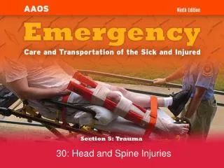
SPINE INJURIES Dr. Waleed Dabbas Consultant neurosurgeon College of Medicine Al Balqa University Al Salt –Jordan March 3, 2019
Definition • Injury to any of following • Bony elements • Soft tissues • Neurological structures • Concerns • Instability of vertebral column • Actual or potential neurological injury soon or later • Development of chronic pain syndrome • Further neurological decline in future
EPIDEMIOLOGY • Most frequently a problem in the young males • 63% age 16-30 • 4:1 male: female • 10:1 Adult to kids • Mechanism • MVA 50% • Falls 21% • Sports-related 14% • “Acts of Violence” 15%
EPIDEMIOLOGY • Multi trauma • Head injuries • Chest injuries • Abdominal injuries • Long bone fractures • Association with Alcohol/Drugs • 20% of patients with major spine injury will have a second spine injury
Pediatrics • Only 5% of SCI occur in children • C –spine is the most vulnerable segment 42%. Of these 67% of C- spine injuries occur in the upper segment • 31% thoracic • 27%lumbar • Fatality rate is higher with pediatric spine injury than with adults. Opposite to head injuries • Neural and soft tissue involvement are common
What happens when the spinal cord is injured? local swelling at the site of injury which pinches off blood (hypoperfusion and ischemia) • Excessive release of glutamate and excitotoxicity of neurons and oligodendrocytes at the site of injury • Infiltration by immune cells (microglia,neutrophils) • Free radical toxicity • Apoptosis/necrosis
EXPERMINTALLY active cell death as well as passive necrosis may mediate damage after injury. After spinal cord injury (SCI) in the rat, typical posttraumatic necrosis occurred, but in addition, apoptotic cells were found from 6 hours to 3 weeks after injury, mainly in the spinal white matter. Apoptotic cells were positive for oligodendrocyte markers. After SCI in monkeys, apoptotic cells were found within remote degenerating fiber tracts. Both secondary degeneration at the site of SCI and the chronic demyelination of tracts away from the injury appear to be due in part to apoptosis. As cytokines mediate oligodendrocyte death, it seems likely that chronic demyelination after CNS injury shares features with chronic degenerative disorders like multiple sclerosis.
biomechanics of spinal injury • Traumatic forces • AXIAL: elements pushed together • DISTRACTION: elements are pulled apart • FLEXION: severe forward bending • EXTENTION: severe backward bending • SHEAR: forces parallel to the surface on which it acts • ROTATIONAL: torsional forces
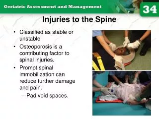
Classification of fractures • Stable: able to carry normal loads • Unstable: unable to carry physical loads • Simple, dislocation, sub laxation, wedge, burst. • 3-COLUMN OF DENIS • Anterior: ALL and anterior 2/3 of V. body and disc • Middle: posterior 1/3 and PLL • Posterior: pedicles, lamina, facets, ligaments
Patterns of neurological injury Following syndromes need to be recognized • Central cord syndrome • Anterior spinal artery syndrome • Brown sequard syndrome • Complete versus incomplete injuries • Conus medullaries injury • Cauda equina syndrome
Complete vs incomplete • It is critical to look for any sign of preserved long tract function of the spinal cord for the purpose of treatment and prognostication. Incomplete Any residual motor or sensory function below the level of injury. look for sacral sparing sensation, voluntary anal contraction, toe flexion, bulbocavernous reflex
Complete vs incomplete • Complete no preservation of any motor or sensory function Bad signs Priapism and abdominal breathing
Neurological level • Motor or Sensory is defined as the lowest level that has completely normal motor or sensory function bilaterally. • C 5 means C6 and below is involved
Spinal shock • Transient physiological disruption which leads to hypotonic areflexic state • Should not impair initial assessment. • Less than an hour • Different from Neurogenic shock When SBP <90 mm hg. Caused by: Loss of muscle tone. Results in venous pooling Sympathetic interruption
Expected outcome • Complete injury unlikely to walk again • About 3% of patients with complete injuries on initial exam will develop some recovery • Complete SCI beyond 24 h indicates no function will recover • Incomplete may make complete or near normal recovery
NEUROLOGICAL STATUS • 25% normal • 20% complete lesion • 55% incomplete lesion/radiculopathy • DETAILED initial assessment imperative • American Spinal Injury Association (ASIA) sheet Pain is the cardinal symptom in alerts Deficits is the main sign in unconscious
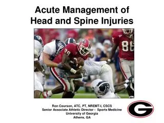
TREATMENT PRINCIPLES • Any patient with significant trauma must be treated as having SCI. • Stabilise • Immobilisation Spinal Cord Injury Surgical Principles • Reduction • Neural decompression • Spinal Stability • Rehabilitation
INITIAL ASSESSMENT • Brief history • Check vitals • Check GCS • Any spine tenderness is crucial • IV access • Bloods • O2 mask • Brief motor exam: ask patient to move arms, hands, legs, and toes
STABILISE • Haemodynamic • Aim oxygen saturation >95% • Systolic BP >90 mm Hg and MAP >80 mm Hg PREVENT SECONDARY INSULT TO CORD • Treat other injuries • NG tube / Ulcer prophylaxis • Bladder care • Temperature • DVT prophylaxis • TEDS / SCD • Clexane
IMMOBILISATION • Hard Collar • Change to Philadelphia / Aspen collar • Traction: tongs, halo • Straight line Spine • No head up (Reverse Trendelenburg) • Log roll patient to turn • Sandbags rigid • Pressure Area Care • Off spinal board ASAP • Rolls • Mattress etc
SPINAL CORD INJURY • Evaluation • Level of injury • Complete / Incomplete • Syndromes • Methyl Prednisolone if within 8 hours of injury • NASCIS III guidelines • 30mg/kg bolus over 15 minutes • Wait 45 minutes • 5.4mg/kg/hr for 23 hours • Continue for additional 24 hours if 3-8 hours past • Relieve Neurological Compression (incomplete) • Controversial
SPINAL STABILITY • Assessment • Immobilisation • Orthosis • Surgical • Reassessment • Treatment
REHABILITATION • Mobilisation • Bowel care • Bladder care • Pain management • Pressure Area Care • Psychology • Placement
IMAGING • Plain radiography • Dynamic radiography ?in certain cases • CT scanning for bony elements • MRI most accurate CT scan and MRI in case of : • clinical suspicion • or abnormal Xray • Myelography ?
PLAIN RADIOGRAPHY • Routine in the ED • AP • Lateral 7 vertebra • Open mouth • (Obliques) • Swimmer’s / Fletcher’s • Visualise Cervicothoracic junction
Dorsal spine X ray: Not accurate • Lumbar Spine Xray: 70% accuracy
PATTERNS OF INJURY • Occipital injuries • Occipitoatlantal dislocation • Occipital condyle fractures • Atalantoaxial (C1/C2) complex injuries • Jefferson’s fractures • Atlantoaxial instability • Rotatory subluxation • Odontoid fractures • Hangman’s fractures • Subaxial Spine
INJURIES INVOLVING OCCIPUT • Occipital-Atlantal dislocation • Rare - ?underestimated • Distraction under hyperflexion • Posterior fusion • Traction considered hazardous • Occipital condyle fractures • Rare • Frequently associated with severe head injury
ATLANTOAXIAL INJURIES • Jefferson’s fracture (C1 arch fracture) • Axial loading • Disruption of C1 ring in multiple sites • Halo immobilisation • May require surgical stabilisation if transverse ligament disrupted
AOD Atlanto-occipital dislocation (AOD) is a devastating condition that frequently results in prehospital cardiorespiratory arrest accounts for 1% of spinal trauma. AOD occurs 3 times more commonly in children than adults, hyperextension. Unstable
ATLANTOAXIAL INJURIES • Atlantoaxial instability • Due to disruption of ligamentous structures, particularly transverse ligament • Also injury to anterior arch of C1, odontoid peg • Tends to fail conservative treatment if ligamentous aetiology • Posterior fixation • Transarticular screws • Wiring techniques
ATLANTOAXIAL INJURIES • Rotatory subluxation • More common in children than adults • Graded I-IV based on integrity of transverse ligament • Often artefactual radiologically
ODONTOID PEG FRACTURES • 7-17% of all cervical spine fractures • Usually present with neck pain • Neurological deficit rare • Type I • Distal odontoid process • Lateral flexion and rotation • Rare usually treated in collar • Type III • Fracture extends into C2 body • Usual heal with Halo / Minnerva
ODONTOID PEG FRACTURES • Type II • Fracture through the synchondrosis of the process with body of C2 • Common (90% of odontoid peg fractures) • Rate of nonunion due to disruption of blood supply • Frequently require surgical fixation • Integrity of transverse ligament important
Posterior C1/2 fusion Anterior screw fixation Maintains rotation Requires real time fluoroscopy SURGERY TYPE II PEG FRACTURES • ss6eedfsddd
HANGMAN’S FRACTURE • Traumatic spondylolisthesis of C2 as a result of bilateral fractures of the C2 pars interarticularis • First reported in 1913 by Wood-Jones • Hyperextension injury • Majority heal in collar
SUBAXIAL CERVICAL SPINE INJURIES • Variety of mechanisms of injury • Three column theory • Anterior column • Anterior longtitudinal ligament • Anterior two thirds of body • Middle column • Posterior third body • Posterior longtitudinal ligament • Posterior column • Pedicles, Laminae • Articular processes • Spinous processes
APPLICATION • Few single column injuries require immobilisation / stabilisation • Most two column injuries usually require immobilisation, some need stabilisation • Almost all three column injuries require immobilisation AND stabilisation
MECHANISMS OF INJURY • Compression – flexion • Compression – extension • Distraction – flexion • Vertical compression • Lateral flexion • Distraction - extension