Download
1 / 51
600 likes | 1.29k Views
Neutron Powder Diffraction Mario Bieringer Department of Chemistry University of Manitoba presented at the 10 th Canadian Neutron Summer School June 15 – 18, 2009 Chalk River, Ontario. Applications of Powder Diffraction. chemistry. physics. engineering. life sciences. biochemistry.

E N D
Neutron Powder Diffraction Mario Bieringer Department of Chemistry University of Manitoba presented at the 10th Canadian Neutron Summer School June 15 – 18, 2009 Chalk River, Ontario
Applications of Powder Diffraction chemistry physics engineering life sciences biochemistry materials science geological sciences archeology
Applications of Powder Diffraction • diffraction = elastic scattering momentum transfer at constant energy, i.e. incident l = diffracted l • Why do we care about diffraction experiments: • identification of materials/phases • determination of structural details, i.e. local environments within an extended structure • evaluation of order and disorder in structures • phase transitions (structural distortions or reconstruction) • chemical reactions • relate structure and properties
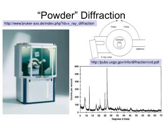
Routine Powder X-ray Diffraction: • Powder X-ray diffractogram • matched with the PDF database • links to crystallographic • data if available (i.e structure • details from ICSD) • semi-quantitative analysis
Routine Powder X-ray Diffraction: Preferred Orientation of flat sample mounts:
In-situ Powder X-ray Diffraction: Intensity (a.u.) 1000 T (0C) 30.0 2q(0) 25 37.0
What is a Powder? • powder = polycrystallinesolidlarge number of crystallites of mm length scale • ideal powders (for diffraction) show random orientation of crystallites • orientational average of single crystals • large number of preparative methods available. • powders can be prepared in large quantities (g, kg, etc.) • fast synthesis • real world materials are often polycrystalline • in reality powders are often multiphasic
Description of Crystallographic Structures • Unit Cell: Smallest repeating unit capable of describing the entire crystal by means of three non-parallel translation vectors. • paralleliped • The 7 Crystal Systems • System u.c. symmetry unit cell parameters • cubic m-3m a = b = c a = b = g = 90˚ • hexagonal 6/mmm a = b ≠ c a = b = 90˚ g = 120˚ • trigonal* 3/mmm a = b ≠ c a = b = 90˚ g = 120˚ • tetragonal 4/mmm a = b ≠ c a = b = g = 90˚ • orthorhombic mmm a ≠ b ≠ c a = b = g = 90˚ • monoclinic 2/m a ≠ b ≠ c a = g = 90˚ b ≠ 90˚ • triclinic -1 a ≠ b ≠ c a ≠ b ≠ g ≠ 90˚ * also described as rhombohedral: a = b = c a = b = g ≠ 90˚
Description of Crystallographic Structures Bravais Lattices:Centering conditions can be described with translation vectors: Lattice Symbol translations lattice points Primitive P +(0, 0, 0) 1 Body centered I +(½, ½, ½) 2 Base centered A +(0, ½, ½) 2 B +(½,0, ½) 2 C +(½, ½, 0) 2 Face centered F +(0, ½, ½), +(½, ½, 0), +(½,0, ½) 4 A total of 14 Bravais Lattices exist (see next page for illustration) cubic: P, I, F orthorhombic: P, I, F, (A,B,C) hexagonal: P monoclinic: P, C trigonal: P triclinic: P tetragonal: P, I
Diffraction Peaks due to Periodicity: Due to the inherent crystal periodicity families of parallel virtual planes can be constructed. The scattering probe diffracts off those planes and generates a pattern that is characteristic of the spacing of the virtual planes and the composition of the unit cell. Virtual planes are identified by the Miller indices (h,k,l). The d-spacings, dhkl, (i.e. the perpendicular distance, between planes) can be determined with Braggs law: l = 2dhkl sinq
Diffraction Peaks due to Periodicity: K2NiF4 structure: tetragonal phase b (001) c a
Powder Diffraction and Symmetry: Predict Bragg positions for primitive cells with V = 64 Å3 (l = 1.54 Å). The higher the symmetry and the smaller the cell volume the smaller the number of diffraction peaks. peak multiplicities decrease from cubic to triclinic: e.g. cubic d(100) = d(-100) = d(010) = d(0-10) = d(001) = d(00-1) orthorhombic d(100) = d(-100) ≠ d(010) = d(0-10) ≠ d(001) = d(00-1)
Description of Crystallographic Structures • Fractional coordinates: • Note: use the crystal system as the coordinate systemx = fraction along a axisy = fraction along b axisz = fraction along c axis • Symmetry Elements: • rotation axes: 1, 2, 3, 4, 6 foldinversion rotation axes: -1, -2, -3, -4, -6 foldmirror planes: mscrew axes (helices): 21, 31, 32, 41, 42, 43, 61, 62, 63, 64, 65glide planes: a, b, c, n, d • Absent reflections due to translations! • 230 Space Groups: (International Tables for Crystallography: Vol. A) • e.g. I41amd (number 141) tetragonal system, body centered 41 screw axis, a glide, mirror, d glide
Description of Crystallographic Structures BaLaMnO4 (K2NiF4 structure type): Space group I4/mmm (# 139) Diffraction pattern: No h+k+l = odd reflections Body centered e (½,½,½) translation) radii =Z Ba(56) = 0,0,0.35 La(57)) = 0,0,0.35 Mn (25) = 0,0,0 O1(8) = 0,½,0 O2(8) = 0,0,1.4
Diffraction Peak Intensities: Structure Factor: X-ray case: Neutron case: Powder Peak Intensities: F(hkl) = structure factorfj = X-ray form factorb = neutron scatt. lengthh,k,l = Miller indicesxj, yj, zj = atomic coordinates of atom jBj = thermal parameterq = diffraction anglel = wavelengthS over entire unit cellI(hkl) = intensityCorrection factors: s = scale factorL = Lorentz-polarization p = multiplicity A = absorption correction P = preferred orientation
Diffraction Peak Intensities: Scattering lengths X-ray form factor, f: Temperature factor, Bj:
Description of Crystallographic Structures BaLaMnO4 (K2NiF4 structure type): b atom=x,y,z Z 5.07fm Ba=0,0,0.35 56e 8.24fm La=0,0,0.35 57e -3.73fm Mn=0,0,0 25e 5.80fm O1=0,½,0 8e 5.80fm O2=0,0,1.4 8e
Peak Widths • coherence length domain sizes crystallite sizes particle sizes • Scherrer equation: • l = wavelength • B = integral breadth • q = diffraction angle • The broader the peaks • the smaller the domains.
direct lattice (crystal structure) reciprocal lattice (diffraction pattern) Diffraction from Randomly Oriented Crystals:
1 single crystal few crystals very large number of microcrystals Debye-Scherrer Camera Diffraction from Randomly Oriented Crystals:
Single Crystal versus Powder Diffraction: • Single crystal diffraction: • Use 3-dimensional reciprocal space: a*, b* and c* axes • Use integrated intensities. • unambiguous peak assignment • Powder diffraction: • Use a large assembly of microcrystals and record an average diffraction pattern in 1-dimension. • Use profile points. ambiguous peak assignment • Reminder: 1/d = 2 sinq/l
Powder diffraction: • Information gets buried in powder average. • Try to extract peak intensities or fit the entire profile. Rietveld Method
Powder X-ray vs. Powder Neutron Diffraction simulations of Pr2Ta2O7Cl2 powder patterns
Simplified Powder Diffractogram Intensity * atomic/ionic positions * temperature factors * order/disorder FWHM * domain sizes * habit 2q (˚) * crystal system * unit cell dimensions * space group
X-ray vs Neutron Powder Diffraction? XRD NPD phase I.D. YES NO indexing YES YES space group YES YES structure refinement YES YES light elements NO YES neighbouring elements NO YES structure solution YES YES
When to Use Neutrons: • Ideally: always (most structure determinations and refinements benefit • from independent measurements (X-ray + neutron)) • In particular: • light and heavy elements present (e.g. oxides, fluoride, hydrides (deuterides) of heavy metals) • neighbouring elements: e.g. Al & Si, Fe & Co, Yb & Lu, Ti & V, etc. • multiple site occupancies: e.g. disordered structures • interest in true bulk properties (i.e averaging over large samples) • complex experimental set ups: chemical reactions, high temperatures, high pressures, low temperatures, magnetic fields, electrochemical cells, chemical/structural processes etc. • magnetic samples • etc.
Are Neutrons going to Answer Your Questions? • look up neutron scattering lengths and cross sections: • http://www.ncnr.nist.gov/resources/n-lengths/ Neutron News, Vol. 3, No. 3, 1992, pp. 29-37
Are Neutrons going to Answer Your Questions? LaFeAs(O1−xFx) LiCoO2, LiNi1-xCoxO2,LiMn2O4 b(La=57) = 8.24 fm b(Li=3) = -1.90 fm b(Fe=26) = 6.58 fm b(Co=27) = 2.49 fm b(As=33) = 9.45 fm b(Ni=28) = 10.3 fm b(O=8) = 5.803 fm b(Mn=25) = -3.73 fm b(F=9) = 5.654 fm b(O=8) = 5.803 fm BiFeO3, BaTiO3, LnMnO3La0.9Ba0.1Ga0.8Mg0.2O2.8 b(Bi=83) = 8.532 fm b(La=57) = 8.24 fm b(Fe 26) = 6.58 fm b(Ba=56) = 5.07 fm b(O=8) = 5.803 fm b(Ga=31) = 7.288 fm b(Mg=12) = 5.375 fm
Two Neutron Powder Diffraction Methods • 1. Continuous Wavelength (Reactor Sources): • single (selectable) wavelength • continuous neutron flux • position sensitive detectors, record neutron rate as a function of diffraction angle • Bragg’s law: l = 2d sinq • Dd/d depends on: - monochromator - take-off angle, - monochromator mosaicity - and sample size • example: C2 http://neutron.nrc-cnrc.gc.ca/c2gen.html
Two Neutron Powder Diffraction Methods 2. Time of Flight Method (Spallation Sources): example: HRPD (ISIS, U.K.) 2 choppers close to the target (6m and 9m)select neutron pulses http://www.isis.rl.ac.uk/crystallography/HRPD/index.htm
Two Neutron Powder Diffraction Methods • 2. Time of Flight Method (Spallation Sources): • white radiation (including epithermal fraction) • pulsed neutron source (50 to 60 Hz) • very high neutron flux during neutron burst • multitude of time of flight detectors, record neutron rate vs. time • Bragg’s law: de Broglie: • mn = neutron mass, vn = neutron velocity, t = flight time, L = flight distance, qd = detector angle • resolution: Dd/d depends on total flight path, L, and detector angle (qd) • use time focusing, large scattering volumes don’t degrade the resolution
Data Comparison 1. Continuous Wavelength (Chalk River – C2 (Pr2Ta2O7Cl2) ): :
Data Comparison 2. Time of Flight Method (IPNS – SEPD (Pr2Ta2O7Cl2) ): d-range: 0.85-10.2Å d-range: 0.33-4.0Å d-range: 0.45-5.4Å
Powder Data Analysis • Record Powder Diffractogram (neutron and/or X-ray) • Identify the phase(s) (X-ray database) • Index phase (X-ray or neutron), i.e. find unit cell (i.e. crystal system) • Identify space group (X-ray or neutron) • If a structural model is available (isostructural compound known) proceed with Rietveld refinement • If no structural model is known proceed with ab-initio structure solution • Consider X-ray and neutron combined analysis • Consider different wavelengths • Consider different temperatures
Rietveld Method • Profile Fitting Method: • use a crystallographic model and compute the corresponding powder diffraction pattern • compare simullation with experimental diffraction data • minimize the difference between experimental and calculated diffraction pattern in a least squares refinement by varying a number of parameters: • minimize: • wi = 1/yi yi = observed intensity yci = calculated intensity • s = scale factor, L(hkl) = Lorentz, polarization and multipicity factorsf(2qi-2q(hkl)) = reflection profile function, P(hkl) = preferred orientation function, A = absorption factor, F(hkl) = structure factor, ybi = background intensity
Rietveld Method Refinable Parameters: GLOBAL PARAMETERS:FOR EACH PHASE zero point atomic positions instrumental profile thermal parameters profile asymmetry site occupancies background parameters scale factor sample displacement lattice parameters absorption preferred orientation crystallite size microstrain magnetic vectors
Rietveld Method • Evaluation of fitting quality (R-factors): • R-pattern, Rp: • R-weighted pattern, Rwp: • R-Bragg factor, RB: • R-expected, Re: Goodness of fit, S:
Rietveld Method A large number of Rietveld programs are available: To a large extent these programs have similar capabilities with individual strengths: Rietveld Programs: GSAS FullProf Rietica Rietan BGMN TOPAS etc. http://www.ccp14.ac.uk/
Rietveld Method Refinement of Pr2Ta2O7Cl2: Parameters:Data: varied: all atomic positions SEPD (IPNS) anisotropic thermal parameters 3 scattering banks unit cell parameters 11005 data points background parameters 60 parameters varied scale factor peak shape parameters Results: Rwp: 0.0448 Rp: 0.0280 Re: 0.0342 S: 1.17
Compare Single Crystal and Powder Results Pr2Ta2O7Cl2: space group: I2/m X-ray single X-tal 1 powder neutron (TOF) 2 atom x y z_________ x y z : Pr 0.6925(1) 0 0.5270(1) 0.69114(24) 0 0.5273(4) Ta 0.0343(0) 0 0.2361(1) 0.03451(13) 0 0.23741(24) Cl 0.8219(3) ½ 0.3891(6) 0.82304(13) ½ 0.39002(25) O1 0.0448(8) ½ 0.2060(18) 0.04401(18) ½ 0.21012(37) O2 0.9190(8) 0 0.0679(16) 0.91882(16) 0 0.07140(34) O3 0.8244(9) 0 0.7519(17) 0.82666(16) 0 0.75746(32) O4 ½ ½ 0 ½ ½ 0 a = 14.109(1) Å a = 14.10763(22) Å b = 3.9247(3) Å b = 3.92548(5) Å c = 6.9051(6) Å c = 6.90610(10) Å b = 92.953(8)0 b = 92.9626(14)0 1 U. Schaffrath and R. Gruhn Naturwissenschaften, 75 140 (1988) 2 P. Baudry and M. Bieringer (2003)
Powder X-ray vs. Neutron Diffraction Li4MgReO6: an order-disorder study
Powder X-ray vs. Neutron Diffraction Li4MgReO6:
High Temperature Studies Neutron diffraction and IN-SITU reactions. Preparation of microwave dielectric oxides of composition Ba3ZnTa2O9 (BZT). 3 BaCO3 + ZnO + Ta2O5 ---- Ba3ZnTa2O9 + 3 CO2
High Temperature Studies Only a high flux neutron diffractometer will permit collection of diffraction patterns within one 1 minute. GEM (ISIS, U.K.) TOF instrument with more than 6000 detectors and medium resolution (18 – 20 m flight path).
* * * * * BZT * =BZT =ZnO =Ta2O5 =BaCO3 Rhombohedral BaCO3and inter- mediate phases T(0C) Starting materials d-spacing(Å) High Temperature Studies 3 BaCO3 + ZnO + Ta2O5 ---- Ba3ZnTa2O9 + CO2
Low Temperature Studies Crystal structure and magnetic long range order in Ba0.05Sr0.95LaMnO4 1 phase Rietveld refinement T=125K phase 1: Ba0.05Sr0.95LaMnO4crystal structure
Mn3+ O2- (Ba2+, Sr2+, La3+) Low Temperature Studies Crystal structure refinement and unit cell evolution in Ba0.05Sr0.95LaMnO4 tetragonal: I4/mmm a = b ≠ c