Download
1 / 12
170 likes | 2.66k Views
Conjunctival Lymphangiectasia: Clinicopathological Features and Surgical Management. Douglas Lyall 1 Fiona Roberts 2 Sathish Srinivasan 1,3 1 Department of Ophthalmology, Ayr Hospital, Ayr, Scotland 2 Department of Pathology, Western Infirmary, Glasgow, Scotland

E N D
Conjunctival Lymphangiectasia:Clinicopathological Features and Surgical Management Douglas Lyall 1 Fiona Roberts 2 Sathish Srinivasan 1,3 1 Department of Ophthalmology, Ayr Hospital, Ayr, Scotland 2 Department of Pathology, Western Infirmary, Glasgow, Scotland 3 University of Glasgow, Glasgow, Scotland The authors have no financial interest in the subject matter of this poster
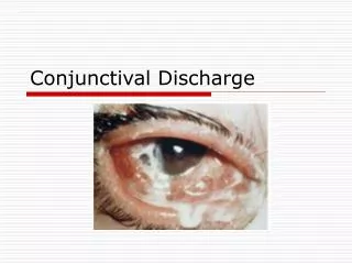
Introduction • Conjunctival Lymphangiectasia (CL) is a localised or diffuse dilatation of the lymphatic channels of the conjunctiva • This gives the clinical appearance of chemosis or of a mobile cyst within the conjunctiva • When localised it may be in response to underlying conjunctival disease • In diffuse cases it may be idiopathic or associated with systemic disease • Patients may complain of symptoms of ocular surface disease including irritation, epiphoria and blurred vision • Previously reported management strategies include simple excision, marsupialization and cryotherapy
Purpose • To report the clinicopathological features of CL • To describe surgical options available in managing this condition Methods • Retrospective case note review of patients with a confirmed histopathological diagnosis of CL • Cases were identified from records of a regional ocular pathology centre • Data recorded: • Age at presentation • Gender • Presenting signs and symptoms • Histopathological features • Management and outcomes
Results • Demographics: • Presenting Characteristics: • Ocular Co-morbidity: Previous Ocular Surgery:
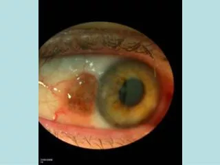
Clinical Appearances Fig. 1 Fig. 2 Fig. 3 Fig. 4 • Inferotemporal translucent swelling of the bulbar conjunctiva in both eyes (arrowed)
Anterior Segment OCT Fig. 5 Fig. 6 • Anterior segment OCT images of both eyes showing cystic spaces within the conjunctiva
Management of Cases • 14 eyes underwent simple excisional biopsy • 2 eyes had excisional biopsy combined with amniotic membrane transplantation (AMT) and fibrin glue • 4 eyes undergoing excisional biopsy had recurrence of chemosis and associated symptoms • 2 eyes undergoing excisional biopsy with AMT had no recurrence of chemosis Outcomes
Surgical Excision with AMT Supplemental Video File
Post Operative Appearance • Fig. 7: Appearances 2 weeks following surgery in the right eye (OD) with AMT. Minimal inflammation with no evidence of wound dehiscence • Fig. 8 & 9: Bilateral absorption of AMT with a well healed conjunctival surface and no recurrence of chemosis at 6 months Fig. 7 Fig. 8 Fig. 9
Histopathology Findings • Squamous metaplasia of the surface mucosa (arrow) • Marked edema of the lamina propria and stroma with numerous dilated lymphatic channels (asterisk) • Chronic inflammation of the stroma including lymphocytes and plasma cells (arrow head) Fig. 10 Fig. 11
Conclusions • Clinical Findings of Conjunctival Lymphangiectasia • Chemosis and hyperaemia occurs in both diffuse and localised forms • It is associated with various ocular surface disease conditions and previous ocular surgery • Histopathological Findings • Diffuse dilatation of conjunctival lymphatic channels and secondary stromal oedema • Associated with secondary chronic conjunctival inflammation
Conclusions • Surgical management options of CL in our series include: • Total excisional biopsy • Total excisional biopsy with amniotic membrane transplantation and fibrin glue • Proposed benefits of performing excision with AMT and fibrin glue: • Eliminates need for suturing and shortens operating time • Reduces postoperative inflammation and discomfort