Download
1 / 28
280 likes | 428 Views
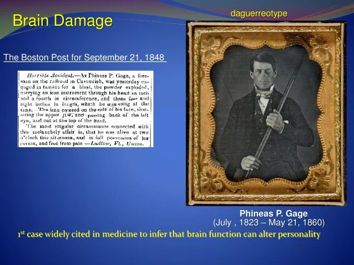
daguerreotype. Brain Damage. The Boston Post for September 21, 1848 . 1 st case widely cited in medicine to infer that brain function can alter personality. Phineas P. Gage. (July , 1823 – May 21, 1860). ~1850. Pattern of injury is very localized -blood vessels spared

E N D
daguerreotype Brain Damage The Boston Post for September 21, 1848 1st case widely cited in medicine to infer that brain function can alter personality Phineas P. Gage (July , 1823 – May 21, 1860)
~1850 Pattern of injury is very localized -blood vessels spared - only frontal cortex damaged - loss of 1 eye Consciousness regained in days - personality/behavior dramatically altered - child-like, profane, disinhibited, inappropriate, inability to control anger and other emotions. Referred to today as classic “hypofrontality” -common with traumatic injury to the frontal lobes Until 2008, commonly thought that he demonstrated these symptoms until his death, 12 years later. But this was not true… “Social Recovery”
Brain Damage • Traumatic brain injury • Strokes • Alzheimer’s Disease • Parkinson’s Disease Other neurological disorders discussed in the book will NOT be subject to test questions
Traumatic Brain Injury FACTS: 1.4 million who sustain a TBI each year in the United States (0.5% of people in U.S.) 50,000 die; 235,000 are hospitalized; and 1.1 million are treated and released from an emergency department. What causes TBI? Falls (28%) Motor vehicle-traffic crashes (20%) Struck by/against (19%) Assaults (11%) Who is at highest risk for TBI? Males are about 1.5 times as likely as females to sustain a TBI.1 The two age groups at highest risk for TBI are 0 to 4 year olds and 15 to 19 year olds Certain military duties (e.g., paratrooper) increase the risk of sustaining a TBI Langlois JA (2006)
Types of Traumatic Brain Injury “Closed Head Injury” Concussion. Concussions are the most common type of TBI. A concussion is damage to nerves or blood vessels in the brain often caused by an impact to the head. Contusions. A contusion is a bruise or bleeding on the brain that can be caused by an impact. Diffuse Axonal Injury. This is tearing of nerve tissue or blood vessels when the brain is jostled in the skull. It can result from shaking (for instance, shaken baby syndrome) or whiplash Coup-countrecoup Injury. impact not only injures the site of impact, but causes the brain to impact with the skull, causing injury to the opposite side of the brain. Symptoms: acute: confusion, visual disturbance, unconsciousness if severe long-term: memory problems, sensory problems . - “dementia pugalistica”
How does traumatic brain injury develop ? • direct, mechanical injury to neurons (axons, dendrites or soma) • damage to blood supply deprives neurons and glia of nutrients • Overactivity of glutamate receptors • Activation of NMDA-type glutamate receptors • Inflow of too much Ca2+into neurons • activates death enzymes that break down neuron • Impairs neuron ability to meet energy requirements • Triggers cell death A little bit of Ca2+ inflow is good at the right time, too much is toxic
Strokes disruption of blood flow in brain 3rd leading cause of death in adults • Hemorrhage: bleeding in brain, blood vessel ruptures • - can be from traumatic injury to blood vessel • - can be from “aneurysm” • weakening of blood vessel wall, balloonlike • dialation forms and can burst • caused by some infections, toxins like cigarette • Smoke. Can also be develomental abnormality.
2. Ischemia: blood flow to a brain region is blocked - a blockage that comes from another part of the body is an “embolism” (vs. thrombosis) for example, a blood clot in leg, can travel to brain and cause ischemia is gets stuck in a smaller artery. “arteriosclerosis”: fat deposits in brain blood vessels grow over time and block blood flow. (can become embolism) How does a loss of blood supply kill neurons ? -deprived neurons become overexcited, release glutamate -glutamate activates NMDA receptors - and because no blood supply=no energy, no reuptake of glutamate
Dementias deterioration of intellectual abilities due to disease *memory, judgment, concentration *usually w/ personality changes, emotional instability What causes dementia ?? 12-15% stroke 12-15% Huntingtons, Parkinson’s Diseases (motor diseases) * other similar disease that are rare • >5% Korsakoff’s syndrome • severe anterogradeamnesia Alzheimer’s Disease (leading cause of severe dementia in U.S.) - +1% of population, most over 65 yrs old
The severity and type of symptoms observed is directly related to death of neurons in several brain regions.
Causes ? We do NOT know what causes Alzheimer’s Disease ! But, we know that…: • Risk of developing it is heritable • there is a “genetic” factor involved 2. Some AD patients have a specific genetic problem That can cause early-onset and severe form (~45 yrs old) 3. But, many other AD patients don’t have family members with AD and don’t have an identified genetic problem Alzheimer’s Disease is probably just 1 term used to describe the symptoms of lots of different diseases that cause brain damage and similar symptoms.
I. Plaques:most AD patients have plaques in parts of the brain that show cell loss in early AD: medial temporal lobe areas (hippocampus) and cortex in late AD: lots of areas (all areas that produce or receive acetylcholine) Key to understanding plaques: • Plaques are made of a protein called beta-amyloid • 2. Beta amyloid come from a normal protein that neurons need to function normally, • called Amyloid Precursor Protein (APP) • 3. but, APP is cut by enzymes into abnormal amounts of beta amyloid. • -neurons can get rid of a small amount of beta amyloid, but • if there is too much, beta amyloid clumps together to form plaques inside of neurons. • Neurons don’t function normally and die-off
II. Tangles:Many AD patients also have “tangles” in neurons Microtubules are long string-like structures that transport things from the cell body to the end of the axon and dendrites Tau proteins (like rungs of a ladder) In AD, these microtubules break apart and collapse into a tangles mess and neurons don’t function normally, so they die-off.
Normal Human Gross Pathology: Ventricles larger gray matter loss white matter loss Human with Alzheimer’s Disease
Drug Treatment of Dementias • Drugs approved by the U.S. Food and Drug Administration • Cognex® • Aricept® • Reminyl® • Exelon® • Namenda® YOU DON’T NEED TO KNOW THESE All are inhibitors of acetylcholinesterase, except Namenda • Acetylcholine is release from “presynaptic” neuron, and then re-absorbed • lots of it in hippocampus and cortex • 2. Acetylcholinesterase is an enzyme that breaks down acetylcholine • 3. That means that more acetylcholine is present in synaptic cleft to stimulate • receptors on neurons in learning/memory areas
Key facts about acetylcholinesterase inhibitors: some improvement in daily living skills some delay in progression of disease BUT, typically a small effect and occurs in a small % of patients Can be very toxic to the liver Namenda® FDA approved to treat late-stage Alzheimer’s the newest approved drug (2004) Namenda® - blocks the NMDA receptor for a very short period of time • Based on theory that over activity of NMDA receptors contribute • to neuron death in Alzheimer’s disease Does it work ?: small effects in a small # of patients But, these small effects have a dramatic effect on quality of life
Parkinson’sDisease mentioned in the Ayurveda, the system of medicine practiced in India as early 7,000 years ago , and in the first Chinese medical text, Nei Jing, which appeared 2500 years ago "An Essay on the Shaking Palsy," published in 1817 by a London physician named James Parkinson,
Symptoms • Difficulty initiating movement • Shuffling gait • “Cogwheel” rigidity • Tremor at rest • Advanced stages may include psychiatric complications • depression • hallucinations • Paranoia • Cognitive decline http://www.youtube.com/watch?v=13ftfmYwfaw
Incidence • Onset usually appears after age of 40 years • but can begin at any time including childhood when it is termed juvenile parkinsonism • affects 1% aged 50 years and over • 10% aged 60 years and over may have undiagnosed, early stages of the disease • about 1½ times more common in men than in women
Subtypes • Primary (idiopathic- most common) • unknown origin but not induced by obvious stimulus • Secondary (parkinsonism) • related to drugs, stroke, or trauma, other stimuli • Familial • genetically linked • accounts for < 20% of the diagnosed cases
Progression: identified too late ? • Considerable dopamine loss must occur before the disease is apparent • clinical diagnosis is usually made after 80% loss in basal ganglia dopamine content • symptoms may emerge after a 60% reduction in basal ganglia dopamine content • The disease is probably present > 20 years before diagnosis
Meso-Striatal Dopamine system COMPONENTS OF THIS SYSTEM • Substantia Nigra- in midbrain • - send fibers to Basal Ganglia • - releases dopamine = movement • Basal Ganglia (part of striatum)- • -sends fibers to motor cortex • -receives fibers from sensory • cortex and substantia Nigra In Parkinson’s Disease Substantia Nigra neurons die = reduced excitatory input into basal ganglia The two parts of the basal ganglia (Caudate and Putamen) also die
Postmortem Analysis brain photographs courtesy of the University of Utah Medical School Roberta J. Seidman, M.D., SUNY Shrunken SubstantiaNigra
PET Scan Loss of cellular activity in the Basal Ganglia
Pathology in Parkinson’s Disease Lewy body pathology: these bodies are circular structures found in cytoplasm. • Dense core of protein called α-synuclein • - normally aids in synaptic vesicle function • Abnormality in gene that makes this protein is a • significant risk for Parkinson’s disease • Many environmental toxins lead to clumping of • α-synuclein too (pesticides, drugs of abuse ?)
Parkin abnormalities ? (familial PD) gene mutation that is a risk factor for Parkinson’s Disease Parkin: a protein found in neurons that transport abnormal proteins to a “proteosome” and tag these proteins with a “death flag” called “ubiquitin”. “Ubiquitination” of a protein signals the proteosome to degrade the protein into its amino acid subparts. Overactivity of Parkin in brain regions controlling motor function can cause over-activation of the proteosome neuronal death
Treatment • Levodopa (L-dopa) therapy (L-dopa is a dopamine precursor) • generally very effective for the first 2 to 5 years of treatment after which the on-off effect develops • Direct-acting dopamine receptor agonists • Experimental brain surgery • lesions of nuclei that inhibit basal ganglia • - remove inhibition of dying neurons, they may be more active = motor control • neural tissue implants (e.g., fetal dopamine cells)
Substantia nigra cells were surgically implanted into the right putamen. Note the continued degeneration seen on the contralateral (left) side revealed by the PET. Recently, Deep Brain stimulation - indwelling electrodes implanted in some of the dying neurons. Patients can stimulate electrodes , provides a brief pulse to these neurons, which rapidly restores some control of motor activity. PET images courtesy of the UCLA School of Medicine, Division of Brain Mapping