Download

1 / 74
740 likes | 760 Views
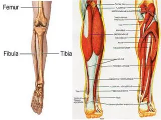
The Leg. Dr. Fadel Naim Orthopedic Surgeon Faculty of Medicine IUG. Fascial Compartments of the Leg. The deep fascia surrounds the leg Continuous above with the deep fascia of the thigh Below the tibial condyles : Attached to the anterior and medial borders of the tibia

E N D
The Leg Dr. Fadel Naim Orthopedic Surgeon Faculty of Medicine IUG
Fascial Compartments of the Leg • The deep fascia surrounds the leg • Continuous above with the deep fascia of the thigh • Below the tibialcondyles: • Attached to the anterior and medial borders of the tibia • It is fused with the periosteum • Two intermuscular septa pass from its deep aspect to be attached to the fibula. • Together with the interosseous membrane, divide the leg into three compartments • Anterior • Lateral • Posterior
INTEROSSEOUS MEMBRANE • A thin but strong membrane connecting the interosseous borders of the tibia and fibula • Most fibers run obliquely downward and laterally • Binds the tibia and fibula together and provides attachment for neighboring muscles • Is continuous below with the interosseous ligament of the inferior tibiofibular joint.
INTEROSSEOUS MEMBRANE • A large opening exists in the upper part of the membrane • Permit the anterior tibial vessels to enter the anterior fascial compartment of the leg • A small opening is present in the lower part of the membrane • For the perforating branch of the peroneal artery to enter the anterior fascial compartment.
RETINACULA OF THE ANKLE • In the region of the ankle joint, the deep fascia is thickened to form a series of retinacula • Keep the long tendons in position and act as modified pulleys.
RETINACULA OF THE ANKLE • The superior extensor retinaculum • A thickened band of deep fascia that is attached to the distal ends of the anterior borders of the fibula and tibia • Near its medial end, it splits to enclose the tendon of the tibialis anterior muscle.
Inferior Extensor Retinaculum • A v-shaped band of deep fascia • Attached by its stem to the upper surface of the anterior part of the calcaneum • The upper limb of the Y is attached to the medial malleolus • The lower limb is continuous with the plantar fascia on the medial border of the foot. • The tendons of: • The tibialis anterior • The extensor hallucis longus • The extensor digitorum longus • The peroneus tertius • Split the upper limb of the retinaculum into superficial and deep layers. • Fibrous bands separate the tendons into compartments each of which is lined by a synovial sheath.
Flexor Retinaculum • A thickened band of deep fascia • Extends from the medial malleolus downward and backward • Attached to the medial surface of the calcaneum • It binds the tendons of the deep muscles to the medial side of the ankle as they pass forward from behind the medial malleolus to enter the sole of the foot. • The tendons lie in compartments each of which is lined by a synovial sheath.
The Superior Peroneal Retinaculum • A thickened band of deep fascia • Extends from the lateral malleolus downward and backward • Attached to the lateral surface of the calcaneum • It binds the tendons of the peroneus longus and brevis to the lateral side of the ankle. • The tendons are provided with a common synovial sheath.
The inferior peroneal retinaculum • A thickened band of deep fascia • Attached to the peroneal tubercle and to the calcaneum • The tendons of peroneus longus and brevis each possess a synovial sheath, which is continuous above with the common sheath.
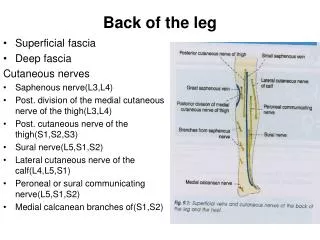
Superficial Veins • Numerous small veins curve around the medial aspect of the leg and ultimately drain into the great saphenous vein
Lymph Vessels • The greater part of the lymph from the skin and superficial facsia on the front of the leg drains upward and medially in vessels that follow the great saphenous vein, to end in the vertical group of superficial inguinal lymph nodes
Lymph Vessels • A small amount of lymph from the upper lateral part of the front of the leg may pass via vessels that accompany the small saphenous vein and drain into the popliteal nodes
CONTENTS OFTHE ANTERIOR FASCIAL COMPARTMENT • Muscles: • The tibialis anterior • Extensor digitorum longus • Peroneus tertius • Extensor hallucis longus. • Blood supply: • Anterior tibial artery. • Nerve supply: • Deep peroneal nerve.
Tibialis Anterior • Origin: • From the upper half of the lateral surface of the tibia • From the interosseous membrane • Insertion: • The tendon passes through both extensor retinacula • Attached to • The medial cuneiform bone • Adjoining base of the first metatarsal bone. • Nerve supply: • Deep peroneal nerve. • Action: • Dorsiflexes the foot at the ankle joint • Inverts the foot at the subtalar and transverse tarsal joints • Assists in holding up the medial longitudinal arch of the foot.
Extensor Digitorum Longus • Origin: • From the upper two thirds of the anterior surface of the fibula • from the interosseous membrane • Insertion: • The tendons pass behind the superior and through the inferior extensor retinacula. • The four tendons then diverge and pass to the lateral four toes. • Nerve supply: • Deep peroneal nerve. • Action: • Extends the toes • Extends the foot at the ankle joint.
Extensor Expansion Of The Foot • On the dorsal surface of each toe, the extensor tendon becomes incorporated into a fascial expansion called the extensor expansion. • The central part of the expansion is inserted into the base of the middle phalanx • The two lateral parts converge to be inserted into the base of the distal phalanx
Peroneus Tertius • Origin: • part of the extensor digitorum longus. • arises from the lower third of the anterior surface of the fibula and the interosseous membrane. • Insertion: • follows the tendons of extensor digitorum longus behind the superior and through the inferior extensor retinacula and shares their synovial sheath. • into the medial side of the dorsal aspect of the base of the fifth metatarsal bone • Nerve supply: • Deep peroneal nerve • Action: • Extends the foot at the ankle joint • Everts the foot at the subtalar and transverse tarsal joints.
Extensor Hallucis Longus • Origin: • From the middle half of the anterior surface of the fibula • From the interosseous membrane • Insertion: • The tendon passes behind the superior and through the inferior extensor retinacula • Into the base of the distal phalanx of the great toe • Nerve supply: • Deep peroneal nerve. • Action: • Extends • Big toe • Foot at the ankle joint • Assists in inversion of the foot at the subtalar and transverse tarsal joints
Artery Of The Anterior Fascial Compartment Of The LegAnterior Tibial Artery • The smaller of the terminal branches of the popliteal artery. • It arises at the level of the lower border of the popliteus muscle • Passes forward into the anterior compartment of the leg through an opening in the upper part of the interosseous membrane • It descends on the anterior surface the interosseous membrane, accompanied by the deep peroneal nerve • In the upper part of its course, it lies deep beneath the muscles of the compartment.
Anterior Tibial Artery • In the lower part of its course, it lies superficial in front of the lower end of the tibia • In front of the ankle joint, the artery becomes the dorsalis pedis artery • passes behind the superior extensor retinaculum • the tendon of the extensor hallucis longus on its medial side • the deep peroneal nerve and the tendons of extensor digitorum longus on its lateral side. • pulsations can easily be felt in the living subject
Branches Anterior Tibial Artery • Muscular branches to neighboring muscles. • Anastomotic branches with branches of other arteries around the knee and ankle joints.
Venae comitantes of the anterior tibial artery • join those of the posterior tibial artery in the popliteal fossa to form the popliteal vein.
Compartment Syndrome • When the pressure within a compartment exceeds the perfusion pressure of the capillaries within that compartment compromising venous blood flow, and limiting capillary perfusion. • Leads to muscle ischemia and necrosis. • TRUE ORTHOPEDIC EMERGENCY
ANTERIOR COMPARTMENT OF THE LEG SYNDROME • Soft tissue injury associated with bone fractures is a common cause • Early diagnosis is critical. • The deep, aching pain in the anterior compartment of the leg that is characteristic of this syndrome • Dorsiflexion of the foot at the ankle joint increases the severity of the pain. • Stretching of the muscles that pass through the compartment by passive plantar flexion of the ankle also increases the pain.
ANTERIOR COMPARTMENT OF THE LEG SYNDROME • In sever cases, the arterial supply is eventually cut off by compression • the dorsalis pedis arterial pulse disappears. • Paralysis of: • The tibialis anterior • the extensor digitorurum longus • the extensor hallucis longus • Loss of sensation in area supplied by the deep peroneal nerve • the skin cleft between the first and second toes. • Decompression by fasciotomy of the anterior compartment of the leg.
Reviewthose“P’s” • Pain? • Paresthesias? • Paralysis? • Pallor? • Pulselessness? • poikilothermia (cool limb)?
CONTENTS OF THE LATERAL FASCIAL COMPARTMENT OF THE LEG • Muscles: • Peroneus longus • peroneus brevis. • Blood supply: • Branches from the peroneal artery. • Nerve supply: • Superficial peroneal nerve
Peroneus Longus • Origin: • From the upper two thirds of the lateral surface of the fibula • Nerve supply: • Superficial peroneal nerve. • Action: • Plantar flexes the foot at the ankle joint • Everts the foot at the subtalar and transverse tarsal joints. • It plays an important part in holding up the lateral longitudinal arch in the foot • serves as a tie to the transverse arch of the foot.
Peroneus Longus • Insertion: • The tendon runs downward behind the lateral malleolus and is held in position by the superior peroneal retinaculum. • The tendon then runs forward on the lateral surface of the calcaneum below the peroneal tubercle. • Here, it is held in place by the inferior peroneal retinaculum. • On reaching the lateral aspect of the cuboid, it winds around the lateral margin and enters a groove on its inferior aspect. • It is inserted into the medial cuneiform and the base of the first metatarsal.
Peroneus Brevis • Origin: • From the lower two thirds of the lateral surface of the fibula • Insertion: • The tendon passes downward behind and directly in contact with the lateral malleolus • held in position by the superior peroneal retinaculum. • The tendon runs forward above the peroneal tubercle of the calcaneum and is held in place by the inferior peroneal retinaculum. • It is inserted into the tubercle on the base of the fifth metatarsal bone. • Nerve supply: • Superficial peroneal nerve. • Action: • Plantar flexes the foot at the ankle joint • Everts the foot at the subtalar and transverse tarsal joints. • It assists in holding up the lateral longitudinal arch of the foot.
TENOSYNOVITIS AND DISLOCATION OF THE PERONEUS LONGUS AND BREVIS TENDONS • Tenosynovitis • Can affect the tendon sheaths of the peroneus longus and brevis muscles as they pass posterior to the lateral malleolus. • Treatment consists of • Immobilization • Heat • Physiotherapy. • Tendon dislocation • The tendons of peroneus longus and brevis dislocate forward from behind the lateral malleolus. • The superior peroneal retinaculum must be torn • It usually occurs in older children and is caused by trauma.
Artery of the lateral Fascial Compartment of the leg • Numerous branches from the peroneal artery which lies in the posterior compartment of the leg, pierce the posterior fascial septum and supply the peroneal muscles.
Superficial Veins • The small saphenous vein • Arises from the lateral part of the dorsal venous arch of the foot • It ascends behind the lateral malleolus in company with the sural nerve. • It follows the lateral border of the tendo calcaneus and then runs up the middle of the back of the leg. • The vein pierces the deep fascia and passes between the two heads of the gastrocnemius muscle in the lower part of the popliteal fossa • Numerous valves along its course.
Superficial Veins • Tributaries • Numerous small veins from the back of the leg. • Communicating veins with the deep veins of the foot. • Important anastomotic branches that run upward and medially and join the great saphenous vein • The mode of termination of the small saphenous vein: • It may join the popliteal vein • It may join the great saphenous vein • It may split in two • One division joining the popliteal • The other joining the great saphenous vein.
Lymph Vessels • Lymph vessels from the skin and superficial fascia on the back of the leg drain upward and either pass forward around the medial side of the leg to • End in the vertical group of superficial inguinal nodes • Drain into the popliteal nodes