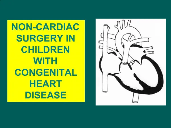
Congenital heart disease (CHD)
Congenital heart disease (CHD). By : - Dr. Sanjeev. Incidence and etiology : -. About 1/100 live births. Sexes are equally affected Higher incidence of PDA and ASD in children born at high altitudes Maternal infection (Rubella) associated with PDA , Pulmonary valve/artery stenosis , ASD.

Congenital heart disease (CHD)
E N D
Presentation Transcript
Congenital heart disease (CHD) By : - Dr. Sanjeev
Incidence and etiology : - • About 1/100 live births. • Sexes are equally affected • Higher incidence of PDA and ASD in children born at high altitudes • Maternal infection (Rubella) associated with PDA , Pulmonary valve/artery stenosis , ASD. • Maternal exposure to drugs and toxins (alcohol) associated with septal defects
Cont… • Congenital heart disease usually manifests in childhood but may pass unrecognized and present in adult life. • Defects which are well tolerated are ASD , may cause no symptoms until adult life or may first be detected incidentally on routine examination or chest radiograph. • Genetic and chromosomal abnormalities like Down’s syndrome cause septal defects and gene defects causing Marfan’s syndrome .
Classification of CHD • Group I : left to right shunts • Group II : right to left shunts • Group III : obstructive lesions
Left to right shunts • ASD • VSD • PDA
Right to left shunts • Fallot`s tetralogy • Tricuspid atresia • Ebstein`s anomaly
Obstructive lesions • Aortic stenosis • Co-arctation of aorta • Valvular regurgitation (AR,MR) • Pulmonary stenosis, • Tricuspid stenosis
Classification of CHD 1. With shunt 2. Without shunt 1. With shunt : • Acynotic : • Ventricular septal defect • Atrial septal defect • Patent ductus arteriosus
Cyanotic • Tetralogy of Fallot • Tricuspid atresia • Transposition of great vessels • Truncus arteriosus • Ebstein anomaly • Pulmonary atresia
2. Without shunt Aortic stenosis • Co-arctation of aorta • Valvular regurgitation (AR,MR) • Pulmonary stenosis • Tricuspid stenosis
Atrial Septal Defect • Abnormal comunication between the two atria • Three types : • 1. Ostium secundum • 2. Ostium primum • 3. sinus venosus
the most common type of ASD - occurs in the center of the septum between the right and left atrium Ostium Secundum Atrial Septal Defect -
next most common type and is located in the lower portion of the atrial septum. will have a mitral valve defect associated with it called a mitral valve cleft. Ostium Primum Atrial Septal Defect
least common type of ASD and is located in the upper portion of the atrial septum Sinus Venosus Atrial Septal Defect
Pathophysiology : - • Physiologically ASD results in leaking of oxygenated blood from the left to right atrium at a minor difference in pressure between two atria -- left to right shunt is thus silent on auscultation -- rt. Atrium receives blood not only through the superior and inferior vena cava but also the blood the blood, shunted from the lt. atrium -- right atrium enlarges in size to accommodate the extra volume of blood -- blood passes through a normal sized tricuspid valve producing a delayed diastolic murmur audible on the bedside at the left sternal border --------------------------------
Cont… • - rt. Ventricle enlarge in size to accommodate the large volume reaching to it -- because of the large volume of blood passing across a normal pulmonary valve a pulmonary ejection murmur is produced and also prolonged ejection phase of the rt. Ventricle -- pulmonary valve, close late and the P2 is delayed -- since rt. Ventricle is fully loaded, further increase in the rt. Ventricle volume during inspiration cannot occur -- second sound is, therefore, widely split and fixed, the P2 is also accentuated
Cont… • The cardiac apex is formed by the enlarged rt. Ventricle and the accentuated P2 is well audible at the apex • Pulmonary artery and its branches enlarge to accommodate the left to right shunt and the lung fields appear plethoric.
Eisenmenger`s syndrome : - • When rt. Ventricular output and pul. blood flow increases -- develop pulmonary hypertension over a period of times -- when pulmonary hypertension is very severe, there may be reversal of blood flow from the right atrium to the left atrium-- such state is then called Eisenmenger`s syndrome
Clinical features : - • Usually asymptomatic in early life. • Increased tendency for respiratory tract infection • Prominent right ventricular pulsation are visible and palpable over the precordium. • Wide and fixed split of the second sound • Murmurs : - • 1. loud ejection systolic murmur is present parasternally over the 2nd and 3rd left intercostal space due to increased blood flow across the pulmonary valve • 2. soft mid diastolic murmur over the apex or over the xiphoid due to increased blood flow across the tricuspid valve
Cont… • Arrhythmias, pulmonary hypertension, Eisenmenger’s syndrome and cardiac failure are the late manifestations. • Cyanosis and clubbing appear when reversal of shunt occurs.
Investigations :- • Electrocardiogram : • Right ventricular hypertrophy • Right axis deviation (ostium secundum) • Left axis deviation (ostium primum) • Echocardiography : • Right ventricular dilatation and hypertrophy • Pulmonary artery dilatation • Doppler : abnormal flow pattern through the defect
Enlargement of right atrium and right ventricle Pulmonary plethora (increased pulmonary vascularity) Chest X ray
Treatment : - • Antibiotic (chest infection) • Small defect -observe • Significant left to right shunting–operate • Operation is contraindicated when there is severe pulmonary HTN and shunt reversal.
Ventricular septal defect : - • Most common congenital heart disease. • 99 % defects lie in the membranous portion.
Pathophysiology :- • Shunting of oxygenated blood from the left to the right -- left ventricle starts contracting before the right ventricle --- the flow of blood from the left ventricle to the right ventricle starts very early in systole and a high pressure gradient is maintained between the two ventricles throughout the systole -- the murmur resulting from the left to right shunt, starts early, masking the first sound and continues throughout the systole with almost the same intensity appearing as a pansystolic murmur on auscultation and palpable as a thrill ----
Cont … • toward the end of systole, the declining left ventricular pressure becomes lower than the aortic pressure -- closure of the aortic valve and occurrence of A2 - left ventricular pressure is still higher than the right ventricular pressure and the left to right shunt continues and the pansystolic murmur masked the first and second sounds.-----
Cont… • -- left to right ventricular shunt occurs during systole at a time when the right ventricle is also contracting and its volume is decreasing -- flow of blood across the normal pulmonary valve results in an ejection systolic murmur at the pulmonary valve -- large volume of blood passing through the lungs is recognized in the chest X – ray as pulmonary plethora --- increased volume of blood finally reaches the left atrium -- left atrial enlargement -- passing through a normal mitral valve the large volume of blood results in a delayed diastolic murmur at the apex ---
Cont… • the intensity and duration of the delayed diastolic murmur depends on the size of the shunt -- the left ventricle has two outlets, the aortic valve allowing the forward flow and the VSD resulting in a back ward leak, it empties relatively early -- results an early A2 -- since the ejection into the right ventricle and pulmonary artery is increased because of the left to right shunt the P2 is delayed ---- second sound is widely split but varies with respiration in patient of VSD with large left to right shunt.
Eisenmenger`s syndrome : - • When the VSD is large, there is icreased pulmonary flow and it may in the long term lead to pulmonary hypertension--- when pulmonary hypertension is very severe and exceeds the systemic pressure, there may be reversal of blood flow from the right ventricle to the left ventricle through the VSD ---such state is then called Eisenmenger`s syndrome
Clinical features : - • Patients with VSD can become symptomatic around 6 – 10 weeks of age. • Palpitation, dyspnea on exertion and frequent chest infection are the main symptoms in older children. • On examination : • Pulse pressure is relatively wide • Precordium is hyperkinetic with a systolic thrill at the left sternal border • Heart size is moderately enlarged • First and second heart sound are masked by a pansystolic murmur at the left sternal border.
Investigations : - • Electrocardiography : - • Depending upon the size of the defect, it may show left ventricular hypertrophy or biventricular hypertrophy. • Echocardiography : - • Enlargement of both right and left ventricle as well as the left atrium • Doppler echocardiography will show the abnormal flow pattern at the site of defect.
Cardiomegaly and Pulmonary plethora Enlargement of main pulmonary artery Large hilar arteries Chest X – ray
Assessment of severity : - Small VSD : • The left to right shunt murmur continues to be pansystolic but the shunt is small , the second sound is normally split and the intensity of P2 is normal • Absence of delay diastolic mitral murmur • If the VSD is very small it acts as a stenotic area resulting in an ejection systolic murmur also known as functional systolic murmur in children which disappears as they grow up because of the spontaneous closure of small VSD.
Large VSD • Results in transmission of left ventricular systolic pressure to the right ventricle -- right ventricular pressure increases and the difference in the systolic pressure in two ventricle decreases -- left to right shunt murmur becomes shorter and softer and on the bed side appears as an ejection systolic murmur • If there is right ventricular outflow obstruction due to pulmonic stenosis, the right ventricular pressure increases and the VSD murmur becomes an ejection systolic murmur
Treatment • Medical management : • Control of CCF • Treatment of repeated chest infections/ prevention and treatment of anemia and infective endocarditis • Surgical treatment (ventriculotomy): indications • 1. CCF occurs in infancy and is not responding to medical management • 2. the left to right shunt is large • 3. If there is pulmonary stenosis , pulmonary hypertension or aortic regurgitation
Patent ductus arteriousus : - • It is a communication between the pulmonary artery and the aorta. • The aortic attachment of the ductus arteriosus is just distal to the left subclavian artery. • It closes functionally and anatomically soon after birth. • Persistence of ductus arteriosus is called patent ductus arteriosus . • Incidence : 11 %
Pathophysiology : - • PDA results in a left to right shunt from the aorta to the pulmonary artery -- the flow occurs both during systole and diastole as a pressure gradient is present throughout the cardiac cycle between two great arteries -- flow of blood results in a murmur which starts in the systole, after the first sound, and reaches a peak at the second sound then diminishes in intensity and is audible during only a part of the diastole --- thus it is a continuous murmur.
Cont… • PDA results in a systolic as well as diastolic overloading of the pulmonary artery -- the increased flow after passing through the lungs reaches the left atrium -- to accommodate the flow the left atrum enlarges in the size -- the increased volume of blood reaching the left atrium enters the left ventricle in diastole, across a normal mitral valve -- increased flow across the mitral valve results in an accentuated first sound as well as a mitral delayed diastolic murmur and is directly related to the size of the left to right shunt -----------------------------------------------
Cont… • 1. small shunts : no mitral diastolic murmur • 2. moderate sized shunts : left ventricular third sound is audible due to rapid filling of the ventricle • 3. large shunts : mitral delayed diastolic murmur • -- left ventricle receives the increased volume of blood during diastole -- cause diastolic overloading of the left ventricle causes a prolongation of the left ventricular systole and an increase in the size of the left ventricle to accommodate the extra volume ---> prolonged ventricular systole results in delayed closure of the aortic valve and a late A2
Cont…. • Large left ventricular volume ejected into the aorta results in dilatation of the ascending aorta --- produce in an aortic ejection click, which is audible all over the precordium and precedes the start of the continuous murmur --- large volume of blood from the left ventricle passing through a normal aortic valve results in an aortic ejection systolic murmur.
Eisenmenger`s syndrome : - • Some patient may develop pulmonary hypertension over a period of many years due to high blood flow into the pulmonary circulation ---- if pulmonary HTN is severe, the flow of blood in PDA may reverse from the pulmonary artery to the aorta -- such state is then called Eisenmenger`s syndrome
Clinical features : - • No symptoms until LVF sets in. • Pulse pressure is wide with low diastolic pressure • Apex beat down and out due to left ventricular overload • Continuous murmur • Thrill may be palpable over the site where the murmur is heard best.
Investigations : - • Electrocardiography : normal or left ventricular hypertrophy • Echocardiography : very useful to demonstrate the PDA and the abnormal flow pattern at the site.
Large Heart shadow and pulmonary artery. Dilated aortic knuckle Hilar and lung vessels dilated Chest X ray : -
Assessment of severity : - • Depends on : - • 1. the larger the heart size the larger the left to right shunt • 2. absence of third sound and delayed diastolic murmur indicates a small left to right shunt. Presence of the third sound indicates a moderate shunt • 3. the wider the pulse pressure the larger the shunt.
Treatment : - • Indomethacin or Ibuprofen can be tried in neonates( useful in first week of life to induce closure) • Reversal of shunt-no treatment • In absence of severe pulmonary hypertension, ductus can be surgically ligated or divided. • Operation should be deferred for several months if there is infective endocarditis as the ductus may be oedematous