ST ELEVATION
ST ELEVATION. Learning Objective. Recognize ST segment elevation in conditions other than acute MI. Significance. Unwarranted thrombolytic therapy Unnecessary emergency angiography Unnecessary anxiety (for intern). Case.


ST ELEVATION
E N D
Presentation Transcript
Learning Objective • Recognize ST segment elevation in conditions other than acute MI
Significance • Unwarranted thrombolytic therapy • Unnecessary emergency angiography • Unnecessary anxiety (for intern)
Case • 25y/o healthy male presents with chest pain and the following EKG findings
Example 1: Normal ST elevation • 1 - 3mm elevation in one or more precordial leads in relation to the end of the PR segment (male pattern) • ST segment is concave
Example 2: Early Repolarization • Most commonly the ST-segment elevation is most marked in V4 with a notch at the J point, and the ST segment is concave • T waves are tall and are not inverted
Example 3: T-wave Inversion • This normal variant differs from the early-repolarization pattern in that the T waves are inverted and the ST segment tends to be coved
Example 4: LV Hypertrophy • Deep S wave • QS pattern in leads V1 through V3 • Elevated ST segment is concave in a pt with uncomplicated LV hypertrophy as compared with convex in a pt with acute concomitant MI
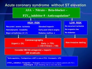
Example 5: Left Bundle Branch Block • Making the dx of acute infarction in the presence of LBBB can be problematic, since the ST segment is either elevated or depressed secondarily, simulating or masking an infarction pattern • Sgarbossa’s criteria is controversial and has not been validated
Example 6: Pericarditis +Myocarditis • ST segment is elevated diffusely in the precordial leads as well as in the limb leads, indicating involvement of more than one coronary vascular territory, which rarely happens in acute myocardial infarction • In addition, the PR segment is depressed, and such depression is the atrial counterpart of ST-segment elevation • ST-segment elevation in patients with acute pericarditisdoes not result in reciprocal ST depression
Example 7: Hyperkalemia • Tall, pointed, and tented T waves • Widened QRS complexes • Low-amplitude or no P waves • Elevated ST segment is often downsloping, a finding that is somewhat unusual in acute myocardial infarction, which is more likely to be characterized by an ST segment that has a plateau or a shoulder or is upsloping
Example 8: Brugada Syndrome • Right bundle-branch block and ST-segment elevation in the right precordial leads in the absence of long QT intervals and any structural heart disease • ST-segment elevation is primarily limited to leads V1 and V2
Case • 25y/o healthy male presents with chest pain and the following EKG findings
Summary • The shape of the ST-segment elevation, the leads involved, other features of the EKG, the clinical setting, and most important, awareness of the conditions that mimic infarction can help differentiate the conditions
References • Kyuhyun Wang, Richard W. Asinger, and Henry J.L. Marriott. ST-Segment Elevation in Conditions Other Than Acute Myocardial Infarction N Engl J Med 2003; 349:2128-2135