Download
1 / 41
420 likes | 532 Views
Learn about the history, purpose, and methods of cardio-pulmonary resuscitation (CPR), including essential steps, equipment, and contraindications. Discover the main stages of resuscitation and how CPR works to save lives.

E N D
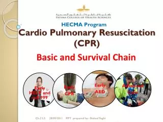
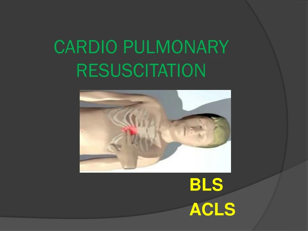
CARDIO PULMONARY RESUSCITATION BLS ACLS
Introduction: Unexpected cardiopulmonary collapse is a medical emergency that requires immediate institution of the artificial measures to support life and to reverse the initiating pathophysiological event. Cerebral resuscitation is the most important goal of advanced cardiac life support. Resuscitation is a continuous process from basic life support (BLS) to advance cardiac life support (ACLS), where BLS initiates the process and ACLS aims to restore and maintain spontaneous respirations and circulations.
DEFINITION: Cardio pulmonary resuscitation (CPR) is a technique of basic life support for the purpose of oxygenation to the heart, lungs and brain until and unless the appropriate medical treatment can come and restore the normal cardiopulmonary function. Cardio pulmonary resuscitation is a series of steps used to establish artificial ventilation and circulation in the patient who is not breathing and has no pulse.
Historical review: • 5000- First artificial mouth to mouth respiration. • 3000 BC- Ventilation. • 1780-First attempt of newborn resuscitation by blowing. • 1874- First experimental cardiac massage. • 1901- First successful direct cardiac massage in man. • 1946- First experimental indirect cardiac massage and defibrillation. • 1960- Indirect cardiac massage. • 1980- Development of cardio pulmonary resuscitation due to works of peter safar.
HOW CPR WORKS: The air we breathe in, travels to our lungs were oxygen is picked up by our blood and then pumped by the heart to our tissue and organs. When a person experiences cardiac arrest-whether due to heart failure in adults or the elderly or an injury such as near drowning, or severe trauma in a child-the heart goes from a normal arrhythmic pattern called ventricular fibrillation, and eventually ceases to beat altogether. This prevents oxygen from circulating throughout the body, rapidly killing cells and tissue. Inessence, cardio (heart) pulmonary(lung) resuscitation (revive, revitalize) serves as an artificial heartbeat and an artificial respirator. CPR may not save the victim even when performed properly, but if started within 4 minute of cardiac arrest and defibrillation is provided within 10 minutes, a person has a 40% chance of survival.
MAIN STAGES OF RESUSCITATION: A (Airway)- ensure open airway by prevention the falling back of tongue, tracheal intubation if possible. B (Breathing) – start artificial ventilation of lung. C (Circulation) – restore the circulation by external cardiac massage. D (Differentiation, drug, defibrillation) – quickly perform differential diagnosis of cardiac arrest; use different medication and electric defibrillation in case of ventricular fibrillation.
CONTRAINDICATIONS Do not resuscitate when a decision not to resuscitate has been noted in chart. This order is often abbreviated to DNR (do not resuscitate), is sometime referred to as no code, and is now discussed with the client on admission and is referred to as an advanced directive.
PURPOSE • Restore cardiopulmonary functioning. • Prevent irreversible brain damage from anoxia.
ASSESSMENT • Determine that the client is unconscious. Shake the client and shout at him or her to confirm if unconscious rather than being asleep, intoxication or hearing impairment. • Assess for the presence of respirations. • Assess carotid artery for pulse.
EQUIPMENTS • A hard flat surface. • No additional equipment is necessary but in hospital setting, an emergency (crash) cart with defibrillator and cardiac monitor should be brought to the bedside. A crash cart contains: • Airway equipment. • Suction equipment. • Intravenous equipment. • Laboratory tubes and syringes. • Pre packed medication for advanced life support.
TYPICALLY THE SEQUENCE OF BLS CONSISTS OF ASSESSMENT AND THE ABCS OF CPR. Assessment It is of crucial importance. It includes • Assess responsiveness by calling the person; shouting and shaking. • Assess breathing by look, listen and feel: Look for chest movements, listen for breath sounds and feel for the movements of the air flow.
BASIC LIFE SUPPORT Airway management Open and clear the airway: This is achieved by head tilt and chin lift maneuver or if there is suspicion/ evidence of head or neck trauma, the jaw thrust maneuver is used.
HEAD TILT CHIN LIFT MANEUVER: Place one hand on the victim’s hairline and place the other hand’s index finger and the middle finger on the chin and apply firm backward pressure.
JAW THRUST MANEUVER: - It is accomplished by placing one hand on each side of the victim’s head, grasping the angles of the victim’s lower jaw, lifting with both hands.
FINGER-SWEEP MANEUVER: - • With the victim’s head up, opens the victim’s mouth by grasping both tongue and the lower jaw between the thumb and fingers and lifting (tongue-jaw lift). • This action draws the tongue from the back of the throat and away from the foreign body. The obstruction may be partially relieved by this maneuver.
If the tongue-jaw lift fails to open the mouth then crossed finger technique may be used. This is accomplished by opening the mouth by crossing the index finger and the thumb and pushing the teeth apart. The index finger of the available hand is inserted along the inside of the cheek and deeply into the throat to the base of the tongue. • A hooking motion is used to dislodge the foreign body and maneuver it into the mouth for removal.
If the tongue-jaw lift fails to open the mouth the crossed finger technique may be used. This is accomplished by opening the mouth by crossing the index finger and the thumb and pushing the teeth apart. The index finger of the available hand is inserted along the inside of the cheek and deeply into the throat to the base of the tongue. • A hooking motion is used to dislodge the foreign body and maneuver it into the mouth for removal.
BREATHING: - After the airway management if the victim is still not breathing, then maintaining head tilt, chin lift positions pinch the nostrils and place the mouth around the victim’s mouth to make a tight seal, take two deep breaths and deliver two positive pressure ventilations; each at least of two seconds duration. When performing mouth-to-mouth ventilation always assess for chest wall movement.
BAG AND MASK VENTILATION • Use a resuscitator bag and mask. • Apply the mask to the victim’s mouth and create a seal by pressing the left thumb on the bridge of the nose and the index finger on the chin. • Use rest of the fingers of the left hand to pull on the chin and the angle of the mandible to maintain the head in extension.
Use the right hand to inflate the lungs by squeezing the bag to its full volume. • Observe the chest wall for symmetric expansion. • Smaller volume (400-600ml) should be attempted during bag and mask ventilation.
CIRCULATION: • The carotid artery is used to determine the absence of pulse. • While maintaining the head tilt position with one hand on the forehead, locate the victim’s trachea with two or three fingers of the other hand, then slides these fingers into the groove between the trachea and the muscles of the neck where the carotid pulse can be felt. • The technique is more easily performed on the side nearest the rescuer. • If on assessment, there are no signs of circulation start external cardiac compressions. • Position hands, arms and shoulders
External cardiac compressions technique consists of serial rhythmic application of pressure on the lower half of the sternum. • The victim is on the horizontal supine position on a flat and hard surface. • The rescuer should be positioned closed to the side of the victim’s chest. • Locate landmark notch hands in the center of the chest, right between the nipples and four fingers above the xiphoid process.
Elbows should be locked and arms are straight. • Rescuer’s shoulders position directly over hands. • Begin compression. • Pressure should come from the shoulders. • Compression should depress victim’s sternum approximately 2 inches. • Don’t allow the fingers to touch the chest wall. • Allow chest to rebound to normal position after each compression.
Perform compression at the rate of 100/min. • Maintain correct position at all times. • Check for signs of circulation every 3-5 min. • Compression: ventilation ratio is 30:2 withonerescuer.Otherwisecontinuecompression. • Exhalation occurs between the two breaths and during the first chest compression of the next cycle. • Perform four complete cycles and then reassess for signs of breathing and circulation.
Five keys aspects to great CPR • Rate • Depth • Release • Ventilation • Uninterrupted
DEFINETION It is asynchronous cardio-version that is used in emergency situation. Defibrillation completely depolarizes the all myocardial cells at once, allowing the sinus node to recapture its role as the pacemaker.
IMPORTANCE OF EARLY DEFIBRILLATIONS • Most frequent arrest frequent arrest rhythm VF/pulselessVT • Treatment is defibrillation. • Successful conversion diminished over time.
NOT USED FOR Sinus rhythm
KEY POINTS TO REMEMBER WHILE DEFIBRILLATING Use a conducting agent between the skins the paddles such as saline pads or electrode paste. This decreases the electrical impedance and helps to prevent burns. • The paddles are placed on the chest wall one the sternal paddle is placed to the right of the sternum, 2’nd intercostals space just below the clavicle. The apex paddle is placed on the left 6’Th intercostals space mid axillary line. • Switch on the defibrillator.
Move the knob of the defibrillator to the required amount of joules. Shock at360 joules. • Exert 20-25 pounds of pressure on each paddle to ensure good skin contact. • Press the charge button. • Call “stand clear” to ensure that personal are not touching the patient or the bed at the time of discharge. • The defibrillator is then discharged by depressing the buttons on the both paddles simultaneously.
GAUGING: • Identify the cause of cardiac arrest by: • Cardiac monitoring. • Lab examination of the blood. HUMAN MENTATION: • Start CPR within 4 min as brain can only survive for four min without oxygen. • Do not interrupt the CPR more than 7min. • Reassess for breathing and circulation every 2-3min.
ECG ECG is the graphical representation of the electrical activity of the cardiac muscles. During CPR the victim’s ECG should be continuously monitored for monitored for monitoring evaluating and recording.
INTENSIVE CARE If the victim’s condition is stable, send the victim to the ICU for close and continuous monitoring. DRUGS THAT CAN BE USED DURING CPR • INJ EPINEPHRIN • INJ ATROPINE • INJ AMIADORONE
TERMINATION OF BASIC LIFE SUPPORT: CPR is stopped as a result of a number of circumstances; these are typically restoration of spontaneous respiration and circulation, complete rescuer exhaustion, or medical decision. Signs of restored ventilation and circulation include: • Struggling movements • Improved color • Return of or strong pulse • Return of systemic blood pressure