Download
1 / 41
470 likes | 1.41k Views
LABORATORY DIAGNOSIS OF BLEEDING DISORDERS. Primary & Secondary Hemostasis Disorders. CIRCULATORY SYSTEM. Low volume, high pressure system Efficient for nutrient delivery to tissues Prone to leakage 2º to endothelial surface damage Small volume loss large decrease in nutrient delivery

E N D
LABORATORY DIAGNOSIS OF BLEEDING DISORDERS Primary & Secondary Hemostasis Disorders
CIRCULATORY SYSTEM • Low volume, high pressure system • Efficient for nutrient delivery to tissues • Prone to leakage 2º to endothelial surface damage • Small volume loss large decrease in nutrient delivery • Minimal extravasation in critical areas irreparable damage/death of organism
HEMOSTATIC DISORDERS • History critical to assessment of presence of disorder • History of bleeding problems in the family • History of spontaneous bleeding • History of heavy menses • History of easy bruising • History of prior blood transfusion • History of prior tooth extractions • History of prior surgery/pregnancy • Physical exam rarely useful except for petechiae or severe hemophiliac arthropathy • Laboratory essential for determining specific defect & monitoring effects of therapy
HEMOSTASISPrimary vs. Secondary vs. Tertiary • Primary Hemostasis • Platelet Plug Formation • Dependent on normal platelet number & function • Secondary Hemostasis • Activation of Clotting Cascade Deposition & Stabilization of Fibrin • Tertiary Hemostasis • Dissolution of Fibrin Clot • Dependent on Plasminogen Activation
COAGULATION TESTINGBasic Testing • Prothrombin Time • Activated partial thromboplastin time (aPTT) • Thrombin Time (Thrombin added to plasma, & time to clot measured) • Fibrinogen • Platelet Count • Bleeding Time
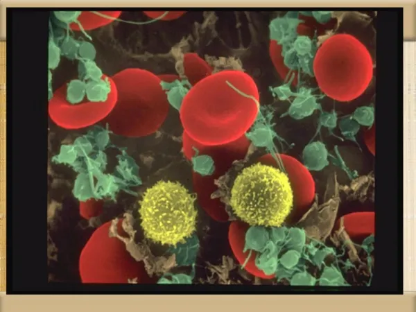
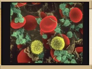
PLATELETS • Anucleate cellular fragmentsMultiple granules, multiple organelles • Synthesis controlled by IL-6, IL-3, IL-11, & thrombopoietin • Circulate as inactive, non-binding concave discs • On stimulation, undergo major shape change • Develop receptors for clotting factors • Develop ability to bind to each other & subendothelium
PLATELET DYSFUNCTIONClinical Features • Mucosal bleeding common • Often see diffuse oozing • Often suspected as a diagnosis of exclusion - ie clotting studies normal, but patient has clinical bleeding disorder • #1 cause of bleeding disorder post-bypass surgery
PLATELET FUNCTION STUDIES • Bleeding Time • Platelet Count • Platelet aggregation studies
BLEEDING TIME • Bioassay • Difficult to standardize • Most reproducible measure of platelet function
PLATELET FUNCTION DEFECTSProlonged Bleeding Time • Congenital • Drugs • Alcohol • Uremia • Hyperglobulinemias • Fibrin/fibrinogen split products • Thrombocythemia • Cardiac Surgery
PLATELET AGGREGATION STUDIES • Multiple agonists used (ADP, epinephrine, collagen, ristocetin) • Add agonists to platelet rich plasma, then measure increase in light transmission as platelets aggregate • Difficult to standardize • Useful for determining cause of platelet dysfunction
PLATELET FUNCTION DEFECTSCongenital • Bernard-Soulier disease (Decreased platelet adhesion • Glanzmann’s thrombasthenia (Decreased platelet aggregation) • γ or δ-storage pool disease (Defective platelet release) • Gray platelet syndrome (Defective platelet release) • Von Willebrand Disease
PLATELET FUNCTION DEFECTSTreatment • Attention to drugs • Platelet transfusion - for bleedingor pre-procedure, esp with congenital defects • Desmopressin (DDAVP) - Shortens bleeding time; ? if decreases bleeding. • Causes release of vWF from endothelial cells • Cryoprecipitate-Same as DDAVP • Dialysis • ? RBC transfusion
THROMBOCYTOPENIACauses-Miscellaneous • Factitious • Macroplatelets • Platelet aggregation • Platelet satellitism • Splenic sequestration • Hemodilution
THROMBOCYTOPENIADecreased production • Decreased megakaryocytes • Normal platelet life span • Good response to platelet transfusion • Neoplastic Causes • Leukemias • Aplastic Anemia • Metastatic Carcinoma • Drugs • Radiotherapy • Primary Marrow Disorders • Megaloblastic Anemias • Myelodysplastic syndromes • Myeloproliferative diseases • Some congenital syndromes
THROMBOCYTOPENIAIncreased Destruction - Causes • Increased megakaryocytes • Shortened platelet life span • Macroplatelets • Poor response to platelet transfusion • Causes • Immune • ITP • Lymphoma • Lupus/rheumatic diseases • Drugs • Consumption • Disseminated intravascular coagulation • Thrombotic thrombocytopenic purpura • Hemolytic/uremic syndrome • Septicemia
COAGULATION CASCADEGeneral Features • Zymogens converted to enzymesby limited proteolysis • Complex formation requiring calcium,phospholipid surface, cofactors • Thrombin converts fibrinogen to fibrin monomer • Fibrin monomer crosslinked to fibrin • Forms "glue" for platelet plug
FVII TF HMWK FIX Ca+2 T Ca+2 or VIIIa/IXa/PL Ca+2 Ca+2 Va/Xa/PL Ca+2 FG F COAGULATION CASCADE INTRINSIC PATHWAY EXTRINSIC PATHWAY FXII FXIIa Surface Active Components FXI Ca+2 VIIa/TF FVIIa FXIa or Ca+2 FIXa VIII VIIIa VIIa/TF FX Middle Components FXa T V Va PT T Common Pathway
FVII TF Ca+2 Ca+2 Va/Xa/PL Ca+2 FG F COAGULATION CASCADE EXTRINSIC PATHWAY ProthrombinTime (PT) FVIIa Ca+2 VIIa/TF FX Middle Components FXa T V Va PT T Common Pathway
HMWK FIX Ca+2 T Ca+2 VIIIa/IXa/PL Ca+2 Ca+2 Va/Xa/PL Ca+2 FG F COAGULATION CASCADE INTRINSIC PATHWAY FXII aPTT FXIIa Surface Active Components FXI FXIa FIXa VIII VIIIa FX Middle Components FXa T V Va PT T Common Pathway
CLOTTING FACTOR DEFICIENCYDetermination of missing factor • Done only if one of screening tests is abnormal • Run panel of assays corresponding to the abnormal screening test, using factor deficient plasmas • PT abnormal - Factors II, V, VII, X • aPTT abnormal - Factors XII, XI, IX, VIII
CLOTTING FACTOR DEFICIENCYDetermination of missing factor • For all but the deficient factor, there will be 50% of normal level of all factors, & clotting assay will be normal • For missing factor, clotting time will be prolonged • If more than one factor level abnormal, implies inhibitor
CLOTTING FACTOR DEFICIENCYCirculating Inhibitor to Clotting Protein • Mixing studies will be abnormal • Need to ensure no heparin is in the specimen • Important to distinguish lupus anticoagulant from circulating anticoagulant to a clotting factor • Former associated with thrombosis • Latter with major hemorrhage • Factor to which inhibitor is directed needs to be determined, along with titer of inhibitor
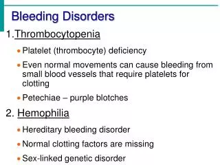
HEMOPHILIA • Sex–linked recessive disease • Disease dates at least to days of Talmud • Incidence: 20/100,000 males • 85% Hemophilia A; 15% Hemophilia B • Clinically indistinguishable except by factor analysis • Genetic lethal without replacement therapy
HEMOPHILIAClinical Severity - Correlates with Factor Level • Mild – > 5% factor level – Bleeding only withsignificant trauma or surgery; only occasionalhemarthroses, with trauma • Moderate – 1–5% factor level – Bleeding with mild trauma; hemarthroses with trauma; occasionally spontaneous hemarthroses • Severe – < 1% factor level – Spontaneous hemarthroses and soft tissue bleeding • Within each kindred, similar severity of disease • Multiple genetic defects • Factor IX > 800 • Factor VIII > 1000
Factor XI Deficiency • 4th most common bleeding disorder • Mostly found in Ashkenazi Jews • Mild bleeding disorder; bleeding mostly seen with procedures/accidents • Levels don’t correlate with bleeding tendency • Most common cause of lawsuits vs. coagulationists
VON WILLEBRAND FACTOR • Large Adhesive Glycoprotein • Polypeptide chain: 220,000 MW • Base structure: Dimer; Can have as many as 20 linked dimers • Multimers linked by disulfide bridges • Synthesized in endothelial cells & megakaryocytes • Constitutive & stimulated secretion • Large multimers stored in Weibel-Palade bodies • Functions:1) Stabilizes Factor VIII2) Essential for platelet adhesion
VON WILLEBRAND DISEASE • Autosomal Dominant Inheritance • Variable Penetrance • 1953 - Patients lack factor VIII • 1957 - Plasma from hemophiliac increase in factor VIII • 1976 - Von Willebrand Antigen discovered • Prevalence: 0.8–1.6% (probable underestimate) • Generally mild bleeding disorder • Variable test results
VON WILLEBRAND DISEASEDiagnostic Studies • aPTT - Prolonged • vWF Activity Level (Ristocetin Cofactor Activity) - Decreased • vWF Antigen Level (“Factor VIII Antigen”) - Decreased • Factor VIII Activity - Decreased • Bleeding Time - Increased • Ristocetin-Induced Platelet Aggregation - Decreased • Multimer Structure - Variable
Initial Therapy of Hemophilia B Modified from Levine, PH. "Clin. Manis. of Hem. A & B", in Hemost. & Thromb., Basic Principles & Practices
VON WILLEBRAND DISEASETherapy • Goal: Correct bleeding time and Factor VIII level • Ideal test for monitoring efficacy of therapy never documented • Treatment usually needed only for surgery or major trauma • DDAVP (Desmopressin - 0.3 μg/kg by infusion • Often effective for Type I; tachyphylaxis develops • Ineffective in Type IIa; relatively contraindicated in Type IIb • MUST TEST FOR EFFICACY PRIOR TO USE • Cryoprecipitate - 1000-1200 units every 12 hours for Types I & II vWD; 2000-2400 units every 12 hours for Type III vWD • Factor VIII concentrate - Do not use, except: • Humate-P (only one containing significant vWF)
CLOTTING FACTOR DEFICIENCYTreatment • For Factor XII & above, no treatment needed • FFP for Factor XI deficiency, factor XIII deficiency • Cryoprecipitate for low fibrinogen, factor XIII deficiency • Factor IX concentrate for deficiency of Vitamin K-dependent clotting factors (important to make sure the one you are using has the factor that you need)
CLOTTING DISORDERSAcquired • Vitamin K deficiency • Liver disease • Coumadin therapy • Heparin therapy • Disseminated Intravascular Coagulation
VITAMIN K DEFICIENCY • Almost always hospitalized patients • Require both malnutrition & decrease in gut flora • PT goes up 1st, 2º to factor VII's short half-life • Treatment: Replacement Vitamin K • Response within 24-48 hours
CLOTTING DISORDERSAcquired • Vitamin K deficiency • Liver disease • Coumadin therapy • Heparin therapy • Disseminated Intravascular Coagulation
LIVER DISEASE • Decreased synthesis, vitamin K dependent proteins • Decreased clearance, activated clotting factors • Increased fibrinolysis 2º to decreased antiplasmin • Dysfibrinogenemia 2º to synthesis of abnormal fibrinogen • Increased fibrin split products • Increased PT, aPTT, TT • Decreased platelets (hypersplenism) • Treatment: Replacement therapy • Reserved for bleeding/procedure