
Visualization of Stroma-Targeted DsRed and Chlorophyll Autofluorescence in Living Mesophyll Cells
This study illustrates the use of stroma-targeted DsRed and chlorophyll autofluorescence for visualizing living mesophyll cells from atg4a4b-1 plants. Fresh leaves from control conditions and IDLs were analyzed immediately after a 5-day treatment. The leaves were treated with 1 μM concanamycin A in a buffered solution before observation. Stromal-targeted DsRed emits green fluorescence while chlorophyll shows red. In merged images, the interaction of DsRed and chlorophyll produces yellow fluorescence, facilitating detailed analysis of mesophyll cell structures.

Visualization of Stroma-Targeted DsRed and Chlorophyll Autofluorescence in Living Mesophyll Cells
E N D
Presentation Transcript
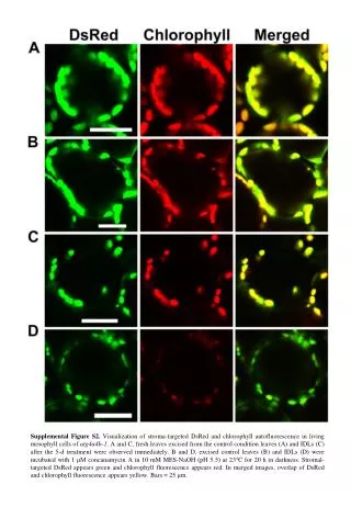
Supplemental Figure S2. Visualization of stroma-targeted DsRed and chlorophyll autofluorescence in living mesophyll cells of atg4a4b-1. A and C, fresh leaves excised from the control condition leaves (A) and IDLs (C) after the 5-d treatment were observed immediately. B and D, excised control leaves (B) and IDLs (D) were incubated with 1 μM concanamycin A in 10 mM MES-NaOH (pH 5.5) at 23C for 20 h in darkness. Stromal-targeted DsRed appears green and chlorophyll fluorescence appears red. In merged images, overlap of DsRed and chlorophyll fluorescence appears yellow. Bars = 25 μm.