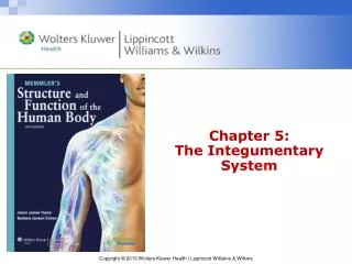
Chapter 5: The Integumentary System
450 likes | 675 Views
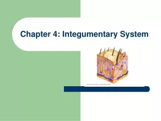
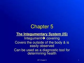
Chapter 5: The Integumentary System. 5.1 Structure of the Skin 5.2 Accessory Structures of the Skin 5.3 Disorders of the Skin 5.4 Effects of Aging 5.5 Homeostasis. 5.1 Structure of the skin. Objectives: Describe the regions of the skin and the hypodermic.

Chapter 5: The Integumentary System
E N D
Presentation Transcript
Chapter 5: The Integumentary System 5.1 Structure of the Skin 5.2 Accessory Structures of the Skin 5.3 Disorders of the Skin 5.4 Effects of Aging 5.5 Homeostasis
5.1 Structure of the skin • Objectives: • Describe the regions of the skin and the hypodermic. • Name two main epidermal layers, and describe their structure and function.
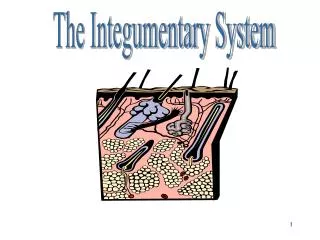
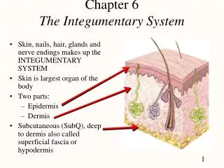
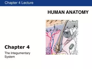
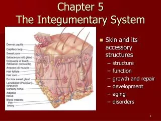
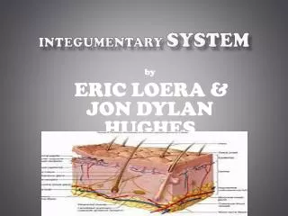
Skin structure • Cutaneous membrane aka the skin. • Skin is an organ system because it has accessory organs. • Two regions: epidermis and the dermis. • Hypodermis: subcutaneous tissue. • * Between skin and muscle.
Epidermis • Outer and thinner region of the skin • Made up of stratified squamous. • Technically five layers.
Epidermis cont… • Epidermis lacks blood vessels!! • Has tightly packed cells. • 80% or more of ‘dust’ is thought to be dead skin!
Stratum basale • Deepest layer of the epidermis. • The farther up they go (away from the dermis) the farther they are from the blood supply. • They eventually die. • Langerhans cells are macrophages found deep in the epidermis. A type of WBC, they phagocytize microbes and then go to the lymphatic system to stimulate an immune response.
Stratum basale • Melanocytes are specialized cells that produce melanin. • This protein makes the skin pigment (of which your teacher has very little!) • The complete inability to produce melanin is albinism. • Carotene is responsible for skin having a yellowish hue. • Hemoglobin in RBCs is what makes Mrs. Holder look pink! • Free nerve endings supply pain and temp. sensations. • Tactile cells signal touching.
Stratum Lucidum • This occurs where constant abrasion has occurred. • Creates calluses. • Found only in thick skin. • Palms of hands, soles of feet, elbows. • These calluses provide protection from constant friction. • Ladies: Ever gotten a pedicure only to have your feet hurt more?
Stratum corneum • The tough, uppermost layer of the epidermis. • Keratin is a fibrous, waterproof protein. • This skin becomes hardened from the keratin. • Minimal on the body, but found on top of the stratum lucidum (where the calluses are!) • Serves as a mechanical barrier against microbe invasion. • Sebaceous glands assist in protecting the skin.
Dermis • Deeper and thicker region • Dense irregular connective tissue • Dermal papillae are fingerlike • projections that are on the upper layer. • They anchor the epidermis and create spirals on fingers that result in ‘fingerprints’. That’s why they are unique and can’t be removed!!!
Dermis cont.. • Contains collagen and elastic fibers. • Collagen fibers are flexible and elastic fibers allow movement of underlying muscles and joints. • Elastic fibers maintain normal skin tension. • Contain blood vessels, supply oxygen and nutrients. • Bedsores result from extended periods of diminished blood flow. • Sensory nerve fibers are here and take nerve impulses to and from the accessory structures.
Hypodermis • Also known as subcutaneous tissue. • Hypodermic needles go into the subcutaneous layer and are called subcutaneous injections. • Loose connective tissue, and adipose tissue. • Fat is an energy storage form that can be called upon when necessary to supply the blood with molecules for cellular respiration. • This is what breaks down when people get old and look wrinkly!!
5.2 Accessory Structures of the Skin • Objectives: • Describe the structure and growth of hair and nails. • Name three glands of the skin, and describe their structure and function.
Hair and Nails • Hair is found on all body parts except palms, soles, lips, nipples, and portions of the external reproductive organs. • Most is fine and downy (peach fuzz). • Hirsutism: condition in women when they produce more male sex hormone than usual and have excessive body and facial hair.
Hair Continued… • Hair follicles: formed from epidermal cells but are located in dermis. • Life spans vary • Eyelash: 3-4 months • Scalp: 3-4 years • Alopecia: hair loss • Androgenic alopecia: male pattern baldness; hereditary • Alopecia areata: patchy hair loss
Hair Continued… • One or more sebaceous glands for each hair • Arrector pili: smooth muscle that connects to follicle • These are goosebumps
Nails • Nail root is the base of the nail where the nail originates from (specialized epithelial cells). • As they grow, they become keratinized. • Cuticle covers the nail root. • Nails grow ~1mm per week • Vascularized dermal tissue accounts for pink color. • White half-moon base, lunula, is due to rapidly reproducing cells in that area.
Glands • Secrete a substance into ducts. • Sweat glands: (sudoriferous glands) are present everywhere • For every square cm, there can be 90 sweat glands on the leg; 400 on the palms and soles. • Apocrine glands open into hair follicles in the anal region, groin, and armpits.
Glands continued… • Eccrine glands open onto the surface of the skin • Homeostasis with perspiration. • Sweat is a form of excretion since it contains water, salts, and urea. • Modified sweat glands in the ear canal, called ceruminous glands, produce earwax.
Sebaceous Glands • Associated with a hair follicle • Secrete oily substance called sebum. • Lubricates hair and skin, waterproofs them, weakens or kills bacteria on skin surface. • If sebum isn’t discharged, then whiteheads/blackheads can occur. • Add bacteria, and you get a pimple. • Acne vulgaris is the most common form of acne.
Mammary Glands • Modified apocrine sweat glands that produce milk only after childbirth.
5.3 Disorders of the Skin • Athlete’s foot: fungal infection that usually involves the skin of the toes and soles. • Use VapoRub…no lie. • Impetigo: contagious, occurs in children, caused by bacterial infection that results in pustules that crust over. • Psoriasis: chronic condition where skin develops pink or reddish patches covered by silvery scales due to overactive cell division.
Disorders Continued… • Eczema: inflammation of the skin • Chemical sensitivity, fabric sensitivity, heat, dryness • Dandruff: caused by an accelerated keratinization in certain areas of the scalp • Urticaria: hives, allergic reaction characterized by the appearance of reddish, elevated patches and often by itcing • Be careful with tattoos…watch out for infection.
Skin Cancer • Melanoma or nonmelanoma • Like all cancers, it’s a mutation of the DNA • Nonmelanoma: include basal cell carcinoma and squamous cell carcinoma • Less likely to metastasize • Basal cell carcinoma: most common type; UV radiation causes a tumor and keeps the immune system from detecting it.
Sqamous cell carcinoma • Begins in epidermis • Five times less common than basal • More likely to spread to nearby organs • Death occurs in 1% of cases. • Excessive UV exposure • May appear to be a wart or scaly growth that bleeds and scabs.
Melanoma • More likely to be malignant • Appearance of an unusual mole • Looks like a spilled ink spot, and can have many shades • Can itch, hurt, or feel numb • Most common in fair-skinned people; especially if they had severe burns as children
Kaposi’s sarcoma • Most commonly seen in patients with AIDS and others whose immune system defenses are weakened or non-functional • Tumors appear as red, blue, or black spots on the skin. • Moles and warts are not usually cancerous. Warts are due to a viral infection.
Wound Healing • Causes an inflammatory response • Characterized by redness, swelling, heat and pain. • If a blood vessel is punctured, it will fill with blood. The blood will then clot, and that clot prevents pathogens and toxins from spreading to other tissues. The clot will dry and harden, becoming a scab. WBCs fight infection, and fibroblasts pull the margins of the wound together.
Wound Healing continued… • Fibroblasts promote tissue regeneration. Basal layer of epidermis produces new cells faster. This brings about scar formation. That scar may or may not be visible from the surface. • Scars contain many collagen fibers that provide maximum strength. • Scar doesn’t contain accessory organs and usually devoid of feeling.
Burns • Mostly heat but can be from radioactive, chemical, or electrical agents. • Two factors affecting severity: depth and extent of burned area • Rule of 9’s • Head and neck is 9%, each arm is 9%, front and back of the trunk is 36% total, each leg is 18%, and the perineum is 1% (anal and urogenital regions).
First-degree Burns • Only epidermis • Redness and pain, but no blisters or swelling • Example: moderate sunburn • Pain subsides within 48-72 hours • Injury heals without further complications or scarring. • Damaged skin peels off in about a week.
Second-degree burn • Extends through the entire epidermis and part of the dermis • Red, pain, AND blistering in the damaged tissue. • Blisters enlarge during the hours after the injury. • Can heal in 10-14 days as long as no infection occurs. • If it’s deeper in the dermis, it could take 30-105 days and scarring is common.
Third-degree burn • Destroy the entire thickness of the skin. • Surface of the wound is leathery and may be brown, tan, black, white, or red. • No pain because the pain receptors have been destroyed. • Blood vessels, sweat glands, sebaceous glands, hair follicles…all been destroyed.
Fourth-degree burn • Down to the bone • Survival is not good unless affected area is very small
Burn becomes critical when… • Second-degree burns cover 25% or more • Third-degree burns cover 10% or more • Any portion of the body has a fourth-degree burn • Third-degree burns occur on the face, hands, or feet. a. Lungs may be damaged from smoke inhalation. b. Loss of joint movement b/c scar tissue formation.
Major Concerns • Fluid Loss • IV of balanced salt solution • Heat Loss • Place in warm environment • Bacterial Infection • Isolation and antibacterial dressings. • Remove tissue and start skin grafting • Autografting: from your own body rather than someone else’s (heterografting)
5.4 Effects of Aging • Thickness is maintained, but mitosis decreases. • Dermis becomes thinner. • Dermal papillae flatten • Epidermis is held less tightly to the dermis • Adipose in hypodermis of face and hands decreases…this is why old people are always cold!!!
Wrinkles • The epidermis is loose. • The fibers are fewer and those remaining are disorganized • The hypodermis has less padding
It sucks to be old… • Hair thins since number of hair follicles decreases. • Fewer sebaceous glands, so the skin tends to crack (lotion!!) • Fewer melanocytes: hair turns gray, skin is paler. • The remaining pigment cells are larger and may appear blotchy • UV damage causes rough skin, mottled pigment, fine lines, wrinkles, skin growths.
5.5 Homeostasis • Objective: List and discuss four functions of the skin that contribute to homeostasis. • 5.4: describe the anatomical and physiological changes that occur in the integumentary system as we age.
Functions of the Skin • Protective Function. Keeps us safe from physical trauma and pathogen invasion. • Regulate water loss. Skin is waterproof and therefore prevents water loss. • Urinary system assist. Perspiration contains salt, ammonia, and urea, so it helps with elimination. • Vitamin D production. UV exposure allows body to convert Vitamin D to calcitriol, which regulates calcium uptake.
More Functions • Sensory information acquisition. Works with CNS to know the external environment. • Regulate body temp. Blood vessels dilate, more blood to the surface, sweat glands become active, sweat takes heat away, and skin cools from it drying. • Cold? The blood vessels contract, which causes shivering, which produces heat.
Hyperthermia and Hypothermia • Hyperthermia: above normal body temp • Hypothermia: below normal body temp • Heat exhaustion: blood pressure may be low, salts may have been lost due to profuse sweating, but the body temp remains high. • Heat stroke: elevated temp with no sweating • Fever: brought on by immune system response • Hypo: shivering, lack of coordination, pulse rate slows, hallucinations, unconsciousness, shallow breathing, rigidity sets in…50% mortality.