Download
1 / 17
170 likes | 368 Views
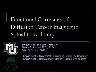
Functional Correlates of Diffusion Tensor Imaging in Spinal Cord Injury. Benjamin M. Ellingson, Ph.D. 1,2 Shekar N. Kurpad, M.D., Ph.D. 2 Brian D. Schmit, Ph.D. 1 1 Department of Biomedical Engineering, Marquette University 2 Department of Neurosurgery, Medical College of Wisconsin.

E N D
Functional Correlates of Diffusion Tensor Imaging in Spinal Cord Injury Benjamin M. Ellingson, Ph.D.1,2 Shekar N. Kurpad, M.D., Ph.D.2 Brian D. Schmit, Ph.D.1 1 Department of Biomedical Engineering, Marquette University 2 Department of Neurosurgery, Medical College of Wisconsin
Motivation • Traditional MRI is not sensitive to axonal injury (Falconer, 1994; Kulkarni, 1988) • Traditional MRI is no better than neurological exam (Flanders, 1999; Shepard, 1999; Bondurant, 1990) • Diffusion Tensor Imaging (DTI) is more sensitive to axon injury (Ford, 1994; Schwartz, 2003) • Objective: Determine if DTI is sensitive to quantitative measures of sensory function (i.e. electrophysiology).
Diffusion Tensor Imaging (DTI) • DTI uses MRI gradients to “tag” diffusing H2O molecules • Apparent Diffusion Coefficient (ADC) is dependent on boundaries to diffusion lADC tADC
Differential Sensitivity of DTI Axonal Damage (Song, 2003; 2002; Nair, 2005; Sun, 2006) ↓ lADC Myelin Damage (Song, 2003; 2002; Nair, 2005; Sun, 2006) ↑ tADC Image Source: Ellingson et al., Concepts in Magn Reson Part A, 2008
Normal SCI Normal Incomplete SCI Complete SCI No temporal Coherence Loss of Amplitude Spinal Somatosensory Evoked Potentials (SpSEPs)
Experimental Spinal Contusion Impactor Vertebral Body
Spinothalamic Tract (STT) & Pain C-fiber input to LSTT (Valeriani, 2007; Li, 1991; Latash, 1988) Ad-fiber input to MSTT (Valeriani, 2007; Latash, 1988) Kandel, 2000, Principles of Neural Science
Hypothesis • Diffusion measurements in the spinothalamic tracts (STTs) correlate with specific components of the SpSEP during high-intensity sciatic nerve stimulation.
Methods - Animals • Neurologically intact (n = 8) • 2 weeks after SCI (n = 8) • 5 weeks after SCI (n = 8) • Spinal Contusion at T8 (Modified from Baker, 2005)
Methods ~ DTI • 9.4-T MR Scanner, Embedded in Agarose Gelatin • 24 axial images though spinal cord (~7 cm) • 6 directions, 100 um resolution • Standard Pulsed Gradient Spin-Echo DTI (PG-SE) • b = 500 s/mm2
Methods ~ SpSEPs • - Animals were anesthetized (Ketamine/Medetomidine IP) • 400 V, 10 mA, 3.5 Hz monophasic square wave, pulse duration 500 us • Amplified 20,000x, sampled at 21 kHz, total of 1000 epochs Image source: Ellingson et al., J Neurotrauma, 2008, Under Review
Results ~ DTI T2-w lADC
Results~Correlation DTI and SpSEPs LSTT lADC Late component (C-fiber) (All animals, R = 0.905, P < 0.001) (2 weeks, R = 0.817, P < 0.01) (5 weeks, R = 0.843, P < 0.01) MSTT lADC Very Early Component (Ad-fiber) (2 weeks, R = 0.812, P < 0.01) (5 weeks, R = 0.841, P < 0.01) Dorsal Columns lADC & tADC Very Early to Early lADC: VE (2 weeks, R = 0.852, P < 0.01) E (5 weeks, R = -0.718, P < 0.05) tADC: VE (2 weeks, R = 0.792, P < 0.01) E (5 weeks, R = 0.835, P < 0.01)
Discussion • LSTT lADC Late component (C-fiber) • MSTT lADC Very Early Component (Ad-fiber) • Dorsal Columns lADC & tADC Very Early to Early
More groups & more specimens Neural stem cells (C17.2) known to cause allodynia Does lADC & SpSEP amplitude increase beyond control? Prognostic capabilities of DTI Does DTI predict final neurological outcome? Motor evoked potentials (MEPs) Is DTI sensitive to motor function deficit? Future Studies
Thank you Brian Schmit, Ph.D. Shekar Kurpad, M.D., Ph.D. Carmen Clark, B.S. James Grosek, B.S. Angie Geiger, B.S. Christy Stadig, B.S. Krishnaj Gourab, M.D. Funding: NIH Falk Foundation Department of Biomedical Engineering, Marquette University Department of Neurosurgery, Radiology, Biophysics at MCW VA Medical Center, Milwaukee WI