Download
1 / 25
250 likes | 375 Views
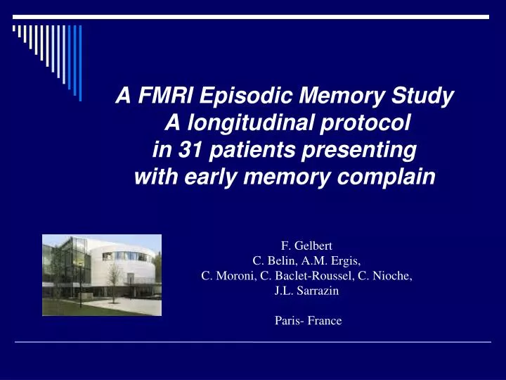
A FMRI Episodic Memory Study A longitudinal protocol in 31 patients presenting with early memory complain. F. Gelbert C. Belin, A.M. Ergis, C. Moroni, C. Baclet-Roussel, C. Nioche, J.L. Sarrazin Paris- France.

E N D
A FMRI Episodic Memory Study A longitudinal protocolin 31 patients presentingwith early memory complain F. Gelbert C. Belin, A.M. Ergis, C. Moroni, C. Baclet-Roussel, C. Nioche, J.L. Sarrazin Paris- France
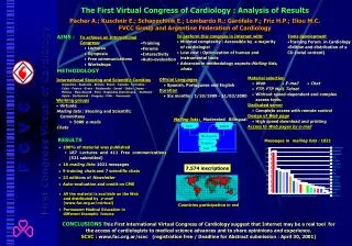
This fMRI study is part of a multimodal protocol including clinical, biological, neuropsychological and standard MR . • The goal of the study was to evaluate the feasability of a FMRI memory protocol in a routine clinical situation
Normal Aging MCI Memory Disorders < 1.5 SD Episodic Memory AD Futur Therapeutic Approaches Early Detection
Longitudinal multimodal study concerning 36 aged patients who complained for Memory Loss 2 FMRI Paradigms Verbal letter fluency is preserved in MCI PARADIGM 1 Memory paradigm PARADIGM 2 Fluency Results to the Fluency test = a baseline standard for each patient
Activation R A R A R A R A Temps R Paradigms • 8 blocks of rest and stimulation periods • Each period : 28 seconds. • During the rest periods, subjects were instructed to think of something unrelated t the stimulation. • Total scanning time: 3 minutes 56 seconds.
Image acquisition • 1.5 Tesla MR scanner : (GE Healhtcare Signa ExHD,) using a phased array 8-elements head coil. • Functional images : T2 EPI/GE. TR : 4.0 ms, TE :60 ms, flip angle 90 , FOV 260 mm, matrix size 64x64, slice thickness 5.0 mm. • Anatomical images : 3D SPGR TR 7.6 ms, TE 1.6 ms, FOV 260 mm, matrix 256x256, NEX 1, slice thickness 1.4 mm.
Memory paradigm from the Memory DSM 48 test (Barbeau et al, Neurology 2002) visual memory task 2 runs Before scanning Visual stimuli were generated using a laptop and a video projector positioned on the scanner bed. The images projected on a white wall were seen by the patients using a mirror adapted to the head coil. more or less than 3 colors. ? silently answer with yes or no.
Verbal Fluency task • Subjects were asked to retrieve as many words as possible with a particular letter defined by an instruction slide P R S T
Recognition task of the Memory test Each target is paired with a distractor on the left or right side of the slide. Instructions are given just before scanning The patient is asked to silently answered Rightor Left for the identified item Right or Left ?
Data analysis and image processing • Statistical Parametric Mapping SPM99 and BAR (home software, HIA Val de Grâce. Paris) • individual T-test maps comparing rest and experimental conditions on a voxel-by-voxel basis. • Data were modeled with a fixed response (box-car)
Results • 36 patients were included in the fMRI protocol • (Age ranged : 70-88, mean age : 78) 19 males, 17 females 14 patients had a normal cognitive status 22 patients presented with MCI • Along the evolution we categorized 3 groups • Cognitively normal individual that remain stable: 12 • MCIpatients that remain stable :7 • MCI patients developing dementia :3
Results • 22 patients have completed the MR protocol at the time being.
Verbal Fluency task Normal subjects right left Strongly lateralised activation in the left hemisphere in the dorsolateral prefontal cortex More bilateralized pattern than younger patients
Normal subjectsMemory test Bilateral activation parietal ,inferior temporal cortex Medial temporal cortex Cingulate cortex Visual areas Middle frontal cortex
Fluency testMCI patients Bilateralized pattern
MCI –AD Convertors Fluency test Initial exam
MCI- AD convertors Memory test Initial exam
Conclusion • We did not faced technical limitations related to the age of the patients. No problem to read the letters and analyse the images The entire procedure was performed within a 30 min examination time Considering the results, the verbal fluency test was reproductively performed even in the early MCI stage. The memory test suffered for the short numbert of patients at the time being. • This experience confirmed the feasability of a Fmri study in a routine clinical practice at least in the first stage of memory loss
And before memory fade away …………….. Special Thanks to the Fondation Philippe Chatrier • Sabine Tranchant, Aurélie Sager • Mrs Gelbert aged 85 who kindly helped us to adapt the different visual parameters to this specific clinical condition