Download

1 / 29
290 likes | 655 Views
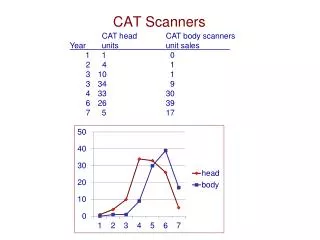
Medical Scanners. Marge Rose 16 th November 2012. Introduction. Confusion – they all look the same CT, MR, SPECT, PET, Ultrasound A plethora of names Why a scan?. Names. CT – computed tomography (was CAT) SPECT – nuclear medicine (was radioisotopes) MR(I) is based on NMR

E N D
Medical Scanners Marge Rose 16th November 2012
Introduction Confusion – they all look the same CT, MR, SPECT, PET, Ultrasound A plethora of names Why a scan?
Names CT – computed tomography (was CAT) SPECT – nuclear medicine (was radioisotopes) MR(I) is based on NMR PET stands for positron emission tomography
Why a scan? An aid to diagnosis Localisation Screening Assessment of function Treatment planning and monitoring Research Reassurance
For each modality We’ll look at • History • Importance • Probe • Signal – few natural ones • Detector – match to signal • What is it detecting?
Ionisation Certain types of radiation can ionise atoms
Ultrasound • Sound is experienced by our ears • Caused by longitudinal pressure waves • We can hear from 20 Hz to 20 kHz • Above 20 kHz - ultrasound
Ultrasound scan 1980
CT scan – uses x-rays They were discovered in 1895 by Röntgen. Here is the very first x-ray – it shows his wife’s hand and was taken in 1895. The first medical use was just a few months later in 1896. X-rays are the most important and widespread of the modalities we will look at in this talk. The method of production is essentially unchanged.
X-ray tube and image But x-ray tubes and images have improved a great deal in over 100 years
The naked CT High Voltage Generator Cooling heat exchanger Cooling oil pump X-Ray tube 120-140kV Detector Array Detector Amplifiers & A/D Converters Collimator
Tomography Patient Grid “Atom” derived from Greek atomos meaning “uncut, indivisible” “Tomography” is from the Greek tomē meaning “cut” or tomos meaning “section” and graphein meaning “to write” X-ray tube Reconstruction of the data by Back projection
CT slice through abdomen Probe 120kV X-ray Interaction Photoelectric, Compton Property X-ray attenuation Image 3D reconstruction from multiple projections
SPECT – uses γ rays Becquerel discovered radioactivity in 1896 The Curies researched into it and Marie opened the first Radium Institute in 1914 Radioisotopes were first used in diagnosis after World War II when radioiodine became readily available Rectilinear scanner appeared in 1951 Anger camera was invented in 1957
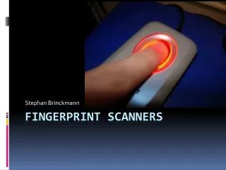
Gamma camera detector First Anger camera I ever saw in use was in 1975 The most common radionuclide used is still Technetium 99m despite supply difficulties Gamma ray energy 140keV Half life of 6 hours ‘No’ beta emission Flexible chemistry
Nuclear Medicine X-ray images show anatomy whereas Nuclear Medicine images show function Uses unsealed radioactive sources introduced into the patient. Patients can still be radioactive when they leave the hospital Gamma cameras are much less common than x-ray machines Very few Nuclear Medicine tests are diagnostic – generally they are highly sensitive but are of low specificity ‘Scans’ can comprise of static or dynamic images, whole body, gated images or SPECT (single photon emission computed tomography)
Whole body imaging A type of static imaging – A whole body bone scan is a very common example
Gated images – the MUGA R-R interval 24 1 2 3 4 Frame or bin
SPECT studies – Myocardial perfusion scan Probe Gamma emitting isotope Interaction Uptake of radiopharmaceutical Property Concentration of pharmaceutical in organ Image • Spatial distribution of counts • SPECT – 3D
Antimatter Each fundamental particle has an antimatter equivalent Same mass but opposite charge Positrons are positive electrons Collide with the first electron they come across to produce annihilation radiation
Positron annihilation 511 keV b+ e.g.18F Coincidence Unit e- 511 keV
PET images Normal Pre-therapy Post-therapy
MR scanner An MR(I) Scanner
MR – souped up NMR If placed in a magnetic field, the nucleus precesses around in the direction of that field Direct in an RF (radiofrequency) pulse and the nucleus can flip to the higher energy state, opposing the field When it relaxes back, it gives off an RF signal which is dependent on the chemical environment • A hydrogen nucleus has spin
MR Probe EM pulses Interaction Resonant energy exchange changes nucleus spin state Property proton density, proton microenvironment Image Map EM signal 3D reconstruction
Artifacts, Hybrid scans The End