Download

1 / 21
220 likes | 425 Views
Spectrophotometers and Concentration Assays. Chapter 7. Learning Outcomes. Describe how a spectrophotometer operates, compare and contrast ultraviolet and visible (white light) spectrophotometers, and give examples of their uses

E N D
Spectrophotometers and Concentration Assays Chapter 7
Learning Outcomes • Describe how a spectrophotometer operates, compare and contrast ultraviolet and visible (white light) spectrophotometers, and give examples of their uses • Determine which type of spectrophotometer is needed for a particular application and the wavelength to be used • Explain the relationship between absorbance and transmittance in spectrophotometry and interpret the meaning of absorbance measurements • Define the term pH and explain the relationship between the concentration of H+ and OH- ions in acids and bases • Describe the proper way to use pH paper and pH meters and which should be used in a specified situation • Justify the need for buffers, describe how buffers are prepared, and calculate the amount of buffering agent needed when making a particular buffer • Explain how protein indicate solutions are used with and without a spectrophotometer • Use a best-fit standard curve to determine the concentration of an unknown protein sample and explain the usefulness of a protein absorption spectrum when trying to isolate a specific protein
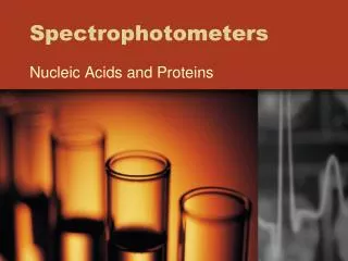
7.1 Using the Spectrophotometer to Detect Molecules Molecules are too tiny to be seen. When an indicator solution changes color, that means a molecule of interest is present.
A VIS spectrophotometer uses white light, composed of the visible spectrum, to detect colorful molecules.
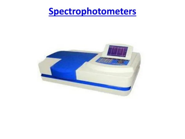
Absorbance, Transmittance, and Reflection. A spectrophotometer measures how light interacts with atoms or molecules in a sample.
Parts of a Spectrophotometer • Lamp • Prism • Sample holder • Display How a Spectrophotometer Works White light hits grating or prism Light is split into colors of the rainbow Wavelength knob directs different colors toward sample
Colors of Light in the Visible Spectrum. Humans can see light with wavelengths of about 350 to 700 nm. How a UV Spectrophotometer Works. Similar to a VIS spectrophotometer, the UV spec shines ultraviolet light or visible light on a sample, and a detector measures the amount of light that passes through, or is absorbed by, the sample.
How Concentration Affects Absorbance. If a sample has twice as many molecules as another, it can absorb twice as much light. This is true at any wavelength. It is important to know a sample’s wavelength of maximum light absorbance, so that the difference in absorbance due to concentration is obvious.
Vocabulary • Ultraviolet light – a wavelength of light that is used to detect colorless molecules • Spectrophotometer – an instrument that measures the amount of light that passes through (is transmitted through) a sample • Nanometer – 10-9 meters; the standard unit used for measuring light • Visible light spectrum – the range of wavelengths of light that humans can see, from approximately 350 to 700 nm; also called white light • Transmittance – the passing of light through a sample • Absorbance – the amount of light absorbed by a sample (the amount of light that does not pass through or reflect off a sample) • Tungsten lamp – a lamp, used for VIS spectrophotometers, that produces white light (350 – 700 nm) • Deuterium lamp – a special lamp used for UV spectrophotometers that produces light in the ultraviolet (UV light) part of the spectrum (200 – 350 nm) • % transmittance – the manner in which a spectrophotometer reports the amount of light that passes through a sample • Absorbance units – (abbreviated “au”) a unit of light absorbance determined by the decrease in the amount of light in a light beam • Absorbance spectrum – a graph of a sample’s absorbance at different wavelengths • Lambdamax– the wavelength that gives the highest absorbance value for a sample
7.1 Review Questions • What is measured in a spectrophotometer? • What is the difference between a UV spectrophotometer and a VIS spectrophotometer? • What happens to the absorbance of a sample as the concentration of a sample increases or decreases? • What color of light has a wavelength of 530 nm? If a molecule absorbs light at 530 nm, what color could it be? What color do we know that it is not?
7.2 Introduction to pH pH is the measure of the level of acid or base in a solution. pH is the measure of the number of hydrogen ions (H+) in a solution. Measuring the pH of a Solution pH paper pH meter Calibrating and Using a pH Meter • Turn on the pH meter. Rinse off the electrode with distilled water. If the meter has a temperature setting knob, set it to room temperature. • Place the electrode in the pH 7 buffer standard. While swirling the solution, adjust the calibration knob until the display reads “7.” • Rinse the electrode with distilled water, being very careful of the very delicate tip. Place the electrode into the solution to be tested. Swirl the solution until the pH display stops changing. Read the pH value. • If the solution is a strong acid or base, use a pH 4 buffer (for the strong acid) or a pH 10 buffer (for the strong base) to slope the meter after it has been calibrated to pH 7.
This pH paper measures the concentration of the H+ ions in a solution.
A pH meter is used to adjust the pH of solutions or to watch the pH of a solution change over time. The pH electrode is sitting in a 50-mL tube of storage solution.
Vocabulary • Acid – a solution that has a pH less than 7 • Base – a solution that has a pH greater than 7 • Hydrogen – a hydrogen atom which has lost an electron (H+) • Neutral – uncharged • Pepsin – an enzyme, found in gastric juice, that works to break down food (protein) in the stomach • pH paper – a piece of paper that has one or more chemical indicators on it and that changes colors depending on the amount of H+ ions in a solution • pH meter – an instrument that uses an electrode to detect the pH of a solution • Buffer standards – solutions, each of specific pH, used to calibrate a pH meter
7.2 Review Questions • If a sample has a pH of 7.8, is it considered an acid, a base or neutral? • What does pH paper measure? • Before a pH meter can be used, it needs to be calibrated. To measure the pH of most solutions, the pH meter is calibrated to what pH? • If the pH of a hot tub is too high, say pH 8, then what should be added to bring it to a neutral pH?
7.3 Buffers Buffer solution resists changes in pH.
7.3 Review Questions • Why must DNA and proteins be stored in buffered solutions? • In what kind of buffer should a DNA sample that was isolated from human cheek cells be stored? • The formula weight of TRIS is 121.14 g/mole. How is 100 mL of 0.02 M TRIS, pH 8.0, prepared?
7.4 Using the Spectrophotometer to Measure Protein Concentration Determining the Wavelength of Maximum Light Absorbance. The lambdamax for this sample is 440 nm. Once lambdamax is determined for a molecule, the spec is set to that wavelength, and all readings for the molecule are made at that wavelength. The lambdamax value is a characteristic of a protein.
Vocabulary • Standard curve – a graph or curve generated from a series of samples of known concentration
7.4 Review Questions • What is lambdamax, and why is it important? • What is the lambdamax for colorless proteins? • How is Bradford reagent used to detect a specific protein in solution? • Which graph is used to determine the concentration of unknown protein samples?