Download
1 / 22
230 likes | 888 Views
Shortened and Internally Rotated Right Leg. SEACSM Case Presentation February 5, 2011 Catherine Rainbow MD Moses Cone Sports Medicine Fellow. Initial History. CC: Right hip pain and inability to move his right leg

E N D
Shortened and Internally Rotated Right Leg SEACSM Case Presentation February 5, 2011 Catherine Rainbow MD Moses Cone Sports Medicine Fellow
Initial History • CC: Right hip pain and inability to move his right leg • 16 year old high school football player who was driven backwards during a hard tackle with his right foot planted while his hip and knee were flexed • Immediate onset of pain in his right hip and was unable to move his right extremity • No changes in sensation of his right leg at the time of injury • Required complete assistance off of the field • Transported via EMS to the local hospital after being on the sidelines for one and a half hours • Past Medical History was significant for an open reduction and internal fixation of his left clavicle. He did not take any regular medications.
Physical Exam • 5’7” and 160 pounds • Gen: Lying on his left side in moderate distress • Musculoskeletal: • Right leg was shortened with the hip flexed, adducted and internally rotated • No tenderness along his spine • His right femoral head was palpable in his buttocks • Muscle spasms were also palpated throughout his thigh and buttocks • He was unable to actively move his right hip and attempts at moving his hip caused severe pain • Range of motion could not be assessed due to pain • He was able to dorsiflex and plantarflex his ankle • 2 + pedal pulses bilaterally • Normal sensation throughout his lower extremities
Panel Questions • Any questions regarding the initial presentation and physical exam?
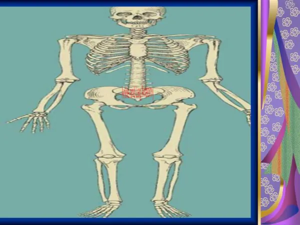
Differential Diagnosis • Fracture of the femur • Acetabular fracture • Pelvic fracture • Dislocated hip • Slipped Capital Femoral Epiphysis (SCFE)
Panel or Audience Questions • Any further questions at this time?
Tests and Results X-ray in the Emergency Department showed a posterior right hip dislocation without signs of a fracture.
Final Diagnosis • Traumatic posterior hip dislocation
Treatment • The patient was diagnosed with a posterior hip dislocation on x-ray in the ED • Orthopedic surgeon was consulted for reduction • Patient was taken to the OR and placed under general anesthesia • The right hip was then carefully flexed, adducted and internally rotated while longitudinal traction was applied to the flexed hip (Bigelow maneuver) • A palpable clunk occurred and concentric reduction of the femoral head was verified on C-arm imaging • Reduction took place approximately 5 hours after the injury occurred
Treatment • The patient stayed in the hospital overnight due to pain and instability with walking on crutches initially • CAT scan of the right hip was performed after the reduction to assess for loose bodies in the joint and fractures
Tests and Results No acute osseous injury of the right hip. No intra-articular loose bodies.
Tests and Results No fractures or loose bodies were identified.
Outcomes • Patient was discharged home the following morning non-weight bearing on crutches • He slept in a hip buttress until pain and strength improved • Followed up by team physician 1 week later with persistent weakness but improved pain • MRI obtained 3 weeks after his injury showed no occult fracture or avascular necrosis but a joint effusion and adductor muscle tears were identified
Outcomes • Four weeks after his injury he started gentle physical therapy and was starting to ambulate without crutches • His strength improved tremendously by two months post closed reduction with formal therapy and was then transitioned to home therapy • He returned to the last football game of the season, three months after his injury • X-rays are to be repeated six months after his initial injury to assess for AVN
Why this is important • 90% of hip dislocations are posterior • Most often occurs in motor vehicle collisions with dashboard injuries • In athletics occurs in collision sports and high speed sports such as football and skiing • Sciatic nerve palsies occur concomitantly 8-20% of the time with a posterior dislocation • Assess neurological exam before and after reduction • Often associated with other injuries such as fracture of the ipsilateral femur or acetabulum and ipsilateral knee injuries • Osteonecrosis of the femoral head is the most common early complication noted in about 10% of patients • Believed to be due to disruption of blood supply to the femoral head via hematoma formation or damage to the junction of the external iliac and common femoral artery or circumflex vessels as seen in cadaver models • Patients need to be followed for 2-3 years with x-rays to assess for AVN • AVN develops in 50% of hips that are dislocated for >12 hours • Other complications include post-traumatic arthritis, femoral nerve injury and chronic pain
Further Questions • Thank you
Judet View To assess the acetabulum
Allis Maneuver Assistant stabilizes the pelvis while physician pulls in-line traction, flexes the hip to 90 degrees and gently alternates between internal and external rotation
Stimson Maneuver Dislocated extremity hang over the side of the table and both knee and hip are flexed to 90 degrees. A downward force is applied to the calf.