Download
1 / 9
90 likes | 224 Views
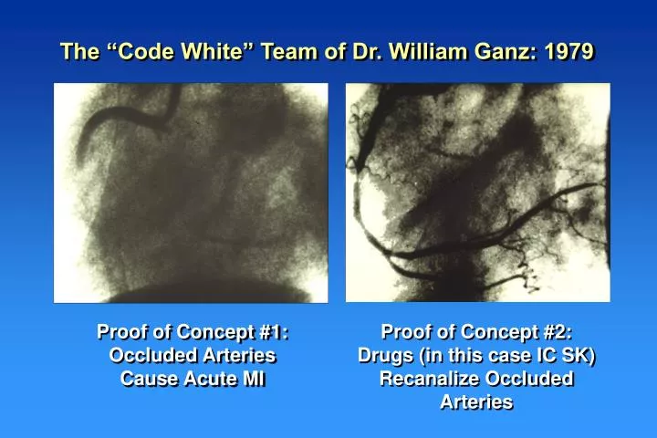
The “Code White” Team of Dr. William Ganz: 1979. Proof of Concept #1: Occluded Arteries Cause Acute MI. Proof of Concept #2: Drugs (in this case IC SK) Recanalize Occluded Arteries. Results of the NIH Sponsored TIMI 1 Trial. % of Patients. % Arteries Opened Documented to be Closed.

E N D
The “Code White” Team of Dr. William Ganz: 1979 Proof of Concept #1: Occluded Arteries Cause Acute MI Proof of Concept #2: Drugs (in this case IC SK) Recanalize Occluded Arteries
Results of the NIH Sponsored TIMI 1 Trial % of Patients % Arteries Opened Documented to be Closed % All Arteries Open at 90 Min. DSMB stopped trial early; felt that greater rate of patency would necessarily translate into improved outcomes Cheesboro et al, Circulation 1987;76: 142-154
The Dark Days of the Open Artery Hypothesis: Results of Early Megatrials Despite the fact that TIMI 1 showed superior patency for tPA over SK, early megatrials showed no difference in mortality Early megatrials used 3 hour dosing of tPA (not front-loaded) and late SQ heparin (not early IV heparin)
Restoration of “Normal” Epicardial Flow Yields Better Outcomes TIMI 1 TIMI 2 TIMI 3 TIMI 0 Occlusion Penetration Slow Flow Normal Flow Unfortunately rate of agreement only 71% 9.3% P=0.003 vs TIMI 0/1 6.1% p<0.0001 vs TIMI 0/1 p<0.0001 vs TIMI 2 % Mortality 3.7% 10 16 33 34 44 4 8 27 13 19 9 15 18 29 34 TIM I 1,4 5,10B Team 2 Team 2 Team 2 TIM I 1,4 5,10B German TIM I 1,4 5,10B German German GUSTO 1 GUSTO 1 GUSTO 1 TAM I 1-7 TAM I 1-7 TAM I 1-7 Sample Size of Pooled Analysis: 5,498 CM Gibson 1998 in Acute Coronary Syndromes
Distal Last Frame First Frame Landmark Definition Definition RCA 1st branch off posterolateral Dye Frame 0: Touches One LCX or No Borders Last branch off most distal OM Frame 21: Dye first enters landmark LAD Dye Frame 1: “Whale’s tail” or “pitchfork” or most distal branch LAD at apex Touches Both Borders & Normal Flow in the Moves Absence of MI : Forward 21.0 + 3.1 frames Gibson, Circulation 1996; 93: 879-888
0 Even Faster Epicardial Coronary Blood Flow is Better 5 6.2% p= 0.003 10 % Risk of In Hospital Mortality 2.8% 15 0.0% (n = 18/640) (n =35/563) 21 (n=41) 14 < CTFC < 40 CTFC < 14 CTFC > 40 “TIMI 4” Flow TIMI 3 Flow Reproducibility: r = 0.97 between readers Accuracy: r=0.88 vs Doppler velocity Hyperemic Flow Gibson, Circulation 1999; 99: 1945-1950
Differences Among the Three Epicardial Arteries Following Thrombolytic Administration in 2,068 TIMI Patients RCA: 90 Min. TIMI 3 Flow: 64.2% Composes 2/3 rds of TIMI 3 flow 90 Min. CTFC: 33.4 Post PTCA CTFC 25.5 Thin walled RV Low filling pressures Distal to Stenosis: 7.8 cm or 59.8% SBP 120.2 mmHg Diameter: 3.23 mm N = 1,044 Percent Stenosis 67.1% Wedge Pressure: 16.4 mm Hg LCX: 90 Min. TIMI 3 Flow: 56.4% 90 Min. CTFC: 40.4 Post PTCA CTFC 36.5 Thick walled LV High filling pressures Distal to Stenosis: 9.2 cm or 70.1% SBP 120.6 mmHg Diameter: 3.10 mm N = 264 Percent Stenosis 69.5% Wedge Pressure: 18.0 mm Hg LAD: 90 Min. TIMI 3 Flow: 46.0% Composes 2/3rds of TIMI 2 flow 90 Min. CTFC: 39.1 Post PTCA CTFC 30.0 Thick walled LV High filling pressures Distal to Stenosis: 11.8 cm or 76.6% SBP 121.7 mmHg Diameter: 2.97 mm N = 778 Percent Stenosis 67.8% Wedge Pressure: 19.7 mm Hg CM Gibson J Am Coll Cardiol 1999; 34: 1403-12