Download
1 / 72
750 likes | 1.2k Views
Adult Health Nursing II Block 7.0. I. Cholecystitis --Acute & Chronic --Cholecystectomy --T-Tubes II. Cancer of Gallbladder III. Pancreatitis --Acute & Chronic IV. Pancreatic Cancer Liver Disease --Cirrhosis --Liver Cancer. Diagnostic Tests.

E N D
Adult Health Nursing II Block 7.0
I. Cholecystitis --Acute & Chronic --Cholecystectomy --T-Tubes II. Cancer of Gallbladder III. Pancreatitis --Acute & Chronic IV. Pancreatic Cancer Liver Disease --Cirrhosis --Liver Cancer Diagnostic Tests TheHepatobiliarySystem Nursing Assessment Nursing Care Planning & Evaluation Block 7.0 Module 5.11
Learning Outcomes • Discuss anatomy and physiology of the hepatobiliary system • Distinguish pathophysiology of hepatobiliary disorders • Discuss clinical manifestations in clients with hepatobiliary disorders • Interpret laboratory findings and diagnostic testing for clients with hepatobiliary disorders • Differentiate interventions and treatment options for clients with hepatobiliary disorders Block 7.0 Module 5.1
Learning Outcomes • Discuss medication management of clients with hepatobiliary disorders • Discuss complications associated with hepatobiliary disorders • Prioritize nursing care for clients with hepatobiliary disorders • Explore teaching plans for clients with hepatobiliary disorders • Differentiate hepatobiliary disorders in the older adult Block 7.0 Module 5.1
Quick A & P Overview Block 7.0 Module 5.1
Quick A & P Overview Block 7.0 Module 5.1
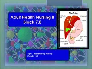
The Biliary System • Comprised of Liver, Gallbladder, and Pancreas • Primary Functions: Secrete enzymes and other substances to promote food digestion • Failure of these organs results in impaired digestion which may result in inadequate nutrition Block 7.0 Module 5.1
Gallbladder Disorders • Function of the Gallbladder: --Collects, concentrates, and stores bile --Releases bile into duodenum when fat is present • Cholecystitis- Acute and Chronic • Cancer of the Gallbladder Block 7.0 Module 5.1
Gallbladder Block 7.0 Module 5.1
Cholecystitis • Inflammation of the Gallbladder • May be acute or chronic • Acute Cholecystitis * 2 Types (Calculous & Acalculous) • Calculous- Chemical Irritation and inflammation resulting from gallstones (cholelithiasis) that obstruct the cystic duct, gallbladder neck, or common bile duct. Exact pathophysiology of gallstones is unknown but cholesterol, bilirubin, calcium, and bile salts play a role in their formation Block 7.0 Module 5.1
Cholecystitis • Acute Cholecytitis • B. Acalculous- Inflammation occurring without gallstones. Biliary stasis due to any condition that affects filling or emptying of the gallbladder Block 7.0 Module 5.1
Cholecystitis • Chronic Cholecysitis • Repeated episodes of cystic obstruction • Calculi are most always present • Gallbladder becomes fibrotic and contracted resulting in decrease motility and deficient absorption Block 7.0 Module 5.1
Cholelithiasis (Gallstones) Block 7.0 Module 5.1
Clinical Manifestations Acute Chronic Jaundice- Yellow discoloration of the skin and mucous membranes due to increased bilirubin in the blood Icterus-Yellow discoloration of the sclera Pruritis- Accumulation of bile salts due to blockage of bile to the duodenum Clay colored stools Steatorrhea- (Fatty Stools) • Sharp pain in right upper quad • Pain with deep inspiration during right subcostal palpation (Murphy’s Sign) • Rebound Tenderness (Blumberg’s Sign) • Nausea, vomiting • Loss of appetite • Fever • Eructation, Flatulence Block 7.0 Module 5.1
Jaundice Block 7.0 Module 5.1
Diagnostics • RUQ Ultrasound- Most diagnostic test • Abdominal X-Rays • Hepatobiliary Scans • Labs: Check your ATI Book • Elevated WBC • Elevated Direct, Indirect, and Total Bilirubin • Elevated AST (with liver dysfunction) • Elevated LDH (with liver dysfunction) • Elevated Serum Cholesterol Block 7.0 Module 5.1
Nursing Interventions and Care • History • Dietary Counseling • - Low fat diet - Promote weight reduction - Avoid gas-forming foods - Small , frequent meals • Pain Management – Meperidine (Demerol) • Antispasmodics • Antiemetics • Prepare for Pre and Post-Op Care Block 7.0 Module 5.1
Cholecystectomy • Removal of the Gallbladder • Nursing Care: • Pain Management • Encourage Splinting to reduce pain • Fundamentals- turn, cough, deep breathe • Monitor incision site • Monitor and record T-Tube drainage-initially bloody then turns to green/brown bile • Assess appetite and response to food Block 7.0 Module 5.1
Cholecystectomy Block 7.0 Module 5.1
T-Tube • Ensures patency of common bile duct • Limits fluid accumulation allowing duct to heal • Surgically placed • Remains up to 6 weeks post-op • Nursing Care of a T-Tube- Iggy pg. 1370, Chart 62-2 Block 7.0 Module 5.1
The Older Adult • May have Diabetes and have atypical presentation of symptoms (absence of pain or fever) • Post Op period is of greater risk • May have difficulty managing care of T-Tube at home • May have difficulty modifying lifelong dietary patterns Block 7.0 Module 5.1
Cancer of the Gallbladder • Rare, more common in women than men • More common in American Indians (etiology unknown) • Most cancers are adenocarcinoma and squamous cell • Begin in inner layer of gallbladder (mucosa) and metastasize to outer organs: Liver, small intestine, and pancreas • Prognosis is poor due to late diagnosis Block 7.0 Module 5.1
Cancer of the Gallbladder Clinical Manifestations Treatment Surgery Radiation Chemotherapy • Similar to cholecystitis • Anorexia, wt loss • Nausea, vomiting • Abdominal Bloating • Fever • Malaise • Jaundice- Advanced • Enlarged liver and spleen • Severe abdominal pain-advanced Block 7.0 Module 5.1
Pancreas • Functions: • Exocrine: secrete enzymes for digestion • Endocrine: Islets of Langerhans cells producing glucagon (Alpha cell) and insulin (beta cell) Block 7.0 Module 5.1
Pancreatitis • Acute- Inflammation of the pancreas resulting from activated pancreatic enzymes autodigesting the pancreas • Mortality can be as high as 20% • Chronic – Progressive destruction of the pancreas with development of calcification and necrosis, possible resulting in hemorrahagic pancreatitis. • Mortality can be as high as 50% Block 7.0 Module 5.1
Acute Pancreatitis • Life threatening inflammatory process • Patho: Premature activation of pancreatic enzymes that destroy ductal tissue and pancreatic cells • Etiology- Mostly unknown however most common causes are: Excessive alcohol intake Biliary tract disease with gallstones Trauma-From surgical procedures Trauma-From diagnostic procedure-ERCP Familial Drug Use Block 7.0 Module 5.1
Acute Pancreatitis Clinical Manifestations Diagnostics Serum Amylase (30-110u/L) Rises within 12-24 hrs, last 4 days Serum Lipase (3-73u/L) Rises slower but last up to 2 weeks Lipase is usually more specific because the pancreas is the only organ that secretes lipase Urine amylase Decrease in serum calcium and magnesium Elevated WBCs Ultrasound and CT • Jaundice • Ascites • Severe, usually sudden onset abdominal/epigastric pain that radiates to back, left flank. Feels “boring” (a feeling like it is going thru the body) • Nausea, Vomiting, Wt. loss • Cullen and Turner signs Block 7.0 Module 5.1
Acute Pancreatitis Cullen’s Sign Turner’s Sign Block 7.0 Module 5.1
Acute Pancreatitis-Complications • Pancreatic infection (most common cause of death) • Hypocalcemia • Hypovolemia • Type I diabetes-Total destruction of the pancreas • Hemorrhage • Septic Shock • Paralytic ileus • ARF • Pneumonia, Atelectasis, Pleural Effusion, ARDS • Coagulation defects- DIC Block 7.0 Module 5.1
Nursing Care • Priority is provide supportive care by relieving symptoms and decreasing inflammation • Always ABCs • Pain Management-Opioids, Dilaudid and MS • IV Fluids, I & O • Rest GI Tract- NPO • Nutrition- TPN • NG Tube • No Smoking • No Alcohol Block 7.0 Module 5.1
Medication Management • Spasmolytics- Pavabid, Cerespan • Anticholinergics- Bentyl- Relieves spasms of muscles in the stomach and intestines , reduces spasms of Sphincter of Oddi (contraindicated in paralytic ileus) • Histamine Receptor Antagonists (HCAs)-Suppress secretion of gastric acid by selectively blocking H2 receptors (Zantac) • Protein Pump Inhibitors (PPIs)- Prilosec • Pancreatic Enzymes- Increases digestion of fats, carbohydrates, and proteins in the GI tract Take immediately before meals with water Viokase, Donnazyme • Antibiotics Block 7.0 Module 5.1
Chronic Pancreatitis • Progressive disease with exacerbations and remissions • Usually develops after repeated episodes of alcohol induced acute pancreatitis • Also caused by chronic obstructive disorders of the common bile duct • Loss of exocrine function-Digestive Enzymes • Loss of endocrine function- Diabetes Block 7.0 Module 5.1
Chronic Pancreatitis Clinical Manifestations Nursing Care Prevent exacerbation Pain Management Pancreatic enzymes Monitor I&O, IV fluids Education: pancreatic enzymes, diet, avoid smoking, caffeine, avoid alcohol, alcohol support groups Comfort measures Insulin therapy Monitor calcium levels • Intense abdominal pain that is continuous, burning or gnawing • LUQ pain- ? Pseudocyst • Steatorrehea or Clay color stools • Weight loss • Exacerbations • Iggy pg. 1378 chart 62-3 Block 7.0 Module 5.1
Pancreatic Cancer • Etiology-Unknown • High incidence in age group 60-80 years • High incidence in smokers • 4th leading cause of cancer death in the U.S. • Spreads rapidly through lymphatic and venous systems to other organs • Vague symptoms usually diagnosed after there is already liver and gallbladder involvement • High mortality rate- Less than 20% live longer than 1 year after diagnosis Block 7.0 Module 5.1
Clinical Manifestations • Jaundice-May be first sign but signifies late stage of disease • Fatigue • Clay colored stools • Dark urine • Abdominal pain-vague • Weight loss, nausea, vomiting • GI bleeding • Anorexia • Splenomegaly- if spleen involved • Hepatomegaly-if liver is involved Block 7.0 Module 5.1
Pancreatic Cancer-Diagnostics • CT scan- visualization of the tumor • Elevated serum amylase and lipase • Elevated carcinoembryonic antigen (CEA) • Elevated alkaline phosphatase and bilirubin • ERCP- Most definitive- Allows for placement of a drain or stent for biliary drainage • Abdominal Paracentesis- Drains fluid and tests for malignant cells Block 7.0 Module 5.1
Endoscopic Retrograde Cholangiopancreatography (ERCP) Block 7.0 Module 5.1
ERCP Block 7.0 Module 5.1
Pancreatic Cancer- Treatments • Management is geared toward preventing tumor spread and decreasing pain. It is palliative not curative • Surgery- Whipple Procedure -Removal of the head of the pancreas, duodenum, parts of the jejunum and stomach, gallbladder, and possibly the spleen. The pancreatic duct is connected to the common bile duct. The stomach is connected to he jejunum • High Risk • Follow with chemotherapy and radiation Block 7.0 Module 5.1
Nursing Care • Post op care- Usually ICU with Whipple Procedure • Pain management • In addition to routine post op care: Assess glucose Assess bowel sounds and stools Assess for infections NPO- NG Tube TPN- Usually started pre-op *Assess for S+S of peritonitis- Board like abdomen *Assess fluids and electrolytes and other labs???? Block 7.0 Module 5.1
Liver Diseases • Function of the Liver is to: • Store • Protect • Metabolize • Cirrhosis • Liver Cancer • Liver Transplants • Hepatitis Block 7.0 Module 5.1
Liver (“Hepato-”) Disorders Block 7.0 Module 5.1
Cirrhosis • Extensive irreversible scarring of the liver caused by a chronic reaction to hepatic inflammation and necrosis Block 7.0 Module 5.1
Cirrhosis Block 7.0 Module 5.1
Cirrhosis Etiology Clinical Manifestations Fatigue Significant change in wt Confusion or difficult thinking GI symptoms and GI Bleeding ABD and liver pain Pruritus Ascites Jaundice and Icterus Petechiae Palmar Erythema Spider Angiomas Fector hepaticus Dependent edema of extremities and sacrum Asterixis READ YOUR ATI BOOK • Alcohol (Laennec’s cirrhosis) • Hepatitis B,C, and D (Post necrotic cirrhosis) • Autoimmune hepatitis • Steatohepatitis-(Fatty Liver Disease) • Drugs and toxins • Biliary disease • Cardiac cirrhosis-(Caused by Heart Failure) Block 7.0 Module 5.1
Ascites Block 7.0 Module 5.1
DiagnosticsCheck Your ATI Book • Serum Liver Enzymes- ALT, AST,ALP- All Increase • Serum Bilirubin- Direct, Indirect, and Total- All Increase • Serum Protein and Serum Albumin- Decrease • CBC- Values Decrease (anemia) • PT/INR- Increase • Serum Ammonia- Increase • Abdominal X-Rays, Ultrasound, CT, MRI • EGD- Detect bleeding or esophageal varices • Liver Biopsy- Most definitive-Measures progression and extent of cirrhosis Block 7.0 Module 5.1
Cirrhosis Secondary to Hepatitis • Acute hepatitis A and acute hepatitis E do not lead to chronic hepatitis. • Acute hepatitis B leads to chronic hepatitis infection in approximately 5% of adult patients. In a few of these patients, the chronic hepatitis B progresses to cirrhosis. • Acute hepatitis D infects individuals already infected by hepatitis B. • Acute hepatitis C becomes chronic in approximately 80% of adults. A minority of these patients (20-30%) will progress to cirrhosis, typically over many years. Block 7.0 Module 5.1
Nursing Care • Comfort Measures • Assess for Bleeding Complications- Blood Transfusions as ordered: Usually RBCs and FFP • Diet and Dietary Teaching High Calorie, Moderate Fat Low-Sodium Low-Protein if encephalopathy and ^ ammonia levels Small-frequent feedings Vitamin Supplements Teach to Avoid Alcohol, May need Referrals Block 7.0 Module 5.1