Download
1 / 28
280 likes | 371 Views
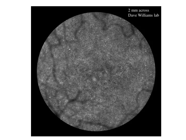
2 mm across Dave Williams lab. Receptive Field Measurements (Kuffler). Receptive Fields: Center-Surround Organization. Harmonic Spatial Stimuli. M = mean level, a = contrast, f = spatial frequency. Receptive Field MTF. Sign-conserving and inverting synapses in the main retinal cell types.

E N D
2 mm across Dave Williams lab
Harmonic Spatial Stimuli M = mean level, a = contrast, f = spatial frequency
Sign-conserving and inverting synapses in the main retinal cell types
Classical Cone Specific Center-Surround Hypothesis(Hubel and Wiesel, 1966; Calkins and Sterling, 2001)
Classical Cone Specific Center-Surround Hypothesis(Hubel and Wiesel, 1966; Calkins and Sterling, 2001)
Anatomy: Midget Cell Surrounds Receive From All Cone Classes(Calkins and Sterling) H1 Horizontals receive non-selective L,M input Amacrine populations receive non-selective L,M input
Hypothesis: Midget Cone Inputs Differ With Eccentricity Central Peripheral
Midget Color Opponency Is Strong In Periphery(Martin, Lee et al., 2001) rg lum
Midget Color Opponency At Various Temporal Modulation Rates(Martin, Lee et al.)
Midget Cell Centers Are Debated(Martin and Lee et al., 2001, Nature)
Methods • In vitro preparation of primate retina: flourescent dye made the parasol, midget, and bistratified cells visible • Was able to target cell by its distinguishing cell body and spatial density • Primary advantage of mounting retina on stage of the microscope: were able to find the ganglion cells easily. • Recordings were taken to show whether midget, parasol, and bistratified cells showed any evidence of S cone input(blue yellow flicker stimuli)
Results • Both midget and parasol cells showed no response to S cone isolating stimuli • The small bistratified cells gave a strong response to S cone isolating stimuli
Conclusions • Isoluminant blue yellow modulation elicited a blue on response in bistratified cells • The physiological differences between M, L, and S cones may reflect the dichotomy between midget cells, parasol cells, and bistratified cells.
Implications of this experiment A Neural circuit may give rise to blue yellow color opponency • Reason: the bistratified cell has a distinct morphology There might be a synaptic pathway for S cone signals formed by bistratified cells and blue cone bipolar cells. Reason: dendritic field of the bistratified cell has the same depth as the axon terminals of the blue cone bipolar cells Other types of ganglion cells might transmit signals from M and L cone opponent cells
From an evolutionary standpoint • Trichromatic vision just recently evolved in mammals • Most mammals have a S cone system.
Questions to Ponder • What about the possible existence of a blue off cell? • What are the advantages of having a blue on pathway? • Why do you think primates only recently evolved trichromatic vision? • What further experiments could be conducted to investigate the question of whether we have a specific circuit for blue yellow opponency?
A Major Retinal Output For S-cone Signals Is the Mosaic of Small Bi-stratifiedGanglion Cells (Calkins)(Calkins and Sterling)
There Is Highly Specialized S-Cone Circuitry (Calkins, J. Del Valle, Kouyama and Marshak, 1992) S-cone S-cone bipolars receive exclusively from S-cones and are pre-synaptic to the small bi-stratified retinal ganglion cells Identified using an antibody for the neuropeptide cholecystokinin (CCK)


![LAB [ 2 ]](https://cdn1.slideserve.com/2335586/lab-2-dt.jpg)