Download
1 / 1
10 likes | 139 Views
Neuronal Coding in the Retina and Fixational Eye Movements. Christian Mendl, Tim Gollisch Max Planck Institute of Neurobiology, Junior Research Group Visual Coding. Overview. Latency emerges as most informative spike response feature Timing reference? (Brain doesn’t know stimulus onset)

E N D
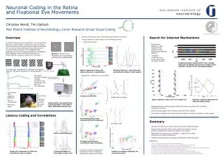
Neuronal Coding in the Retina and Fixational Eye Movements Christian Mendl, Tim Gollisch Max Planck Institute of Neurobiology, Junior Research Group Visual Coding Overview • Latency emerges as most informative spike response feature • Timing reference? (Brain doesn’t know stimulus onset) • → Need several cells Search for Internal Mechanisms So-called “fixational eye movements” are an important feature of normal human vision since they counteract visual perception fading and enhance spatial resolution. Yet it is not yet fully understood how they influence neuronal coding schemes. To investigate these questions, we record the action potential of amphibian retinal ganglion cells, mimicking fixational eye movements by oscillatory shifts of the stimulus. • Relationship between a cell’s receptive field position on the grating and response latency? • → Replace orientations by linear phase shifts for easier analysis Stimuli from Rucci et al., Miniature eye movements enhance fine spatial detail Employing linear phase shifts (color coded). Each pair of ellipses shows the receptive field position relative to the oscillating grating Concrete task: discriminate 5 different orientations based on the spike responses of retinal ganglion cells Spike responses of two cells (blue and green, respectively) Relative latency →time intervals accessible by higher brain regions The upper stimulus modality imitates oscillatory eye movement, and the lower microsaccades • Observation: latencies are correlated • Informative spike response features? • Role of correlations? • Population code? All experiments are performed on Axolotl and Frog (Xenopuslaevis) Latency scatter plot Global drift correction Spike response raster plot for a single cell Relative response latencies for different phase shifts Extracellular recordings from retinal ganglion cells using a MEA (Multi-Electrode-Array) • Response latency matches phase shift and follows reversal of the oscillatory movement direction • Latency range bigger than stimulus movement time interval • First spike is elicited earlier when receptive field moves from a bright to a dark region Latency Coding and Correlations Subtracting global drift reveals internal correlations Summary Shuffling trials • Latency emerges as the most informative spike response feature • Relative spike timings of two cells contain information and are directly accessible to readout by higher brain regions • Responses of cell pairs are correlated → evidence for coding structure via intrinsic interactions • Receptive field position on grating could predict response latency Spike count histogram Compare with latency correlations after shuffling References • Meister et al. (1995), Concerted signaling by retinal ganglion cells. Science 270 • T. Gollisch and M. Meister (2008), Rapid neural coding in the retina with relative spike latencies. Science 319 • S. Martinez-Conde et al. (2006), Microsaccades counteract visual fading during fixation. Neuron 49 • M. Greschner et al. (2002), Retinal ganglion cell synchronization by fixational eye movements improves feature estimation. Nature Neuroscience 5 • M. Rucciet al. (2007), Miniature eyemovementsenhance fine spatialdetail, Nature 447 • M.J. Schnitzer and M. Meister (2003), Multineuronal firing patterns in the signal from eye to brain. Neuron 37 • E. Schneidman et al. (2003), Synergy, redundancy, and independence in population codes. Journal of Neuroscience 23(37) • D.K. Warland et al. (1997), Decoding visual information from a population of retinal ganglion cells. Journal of Neurophysiology 78 Conclusion: there are cell pairs showing internal correlations, additional to global drift effects Single cell responses for different orientations (color coded) Timing histogram of first spike in each trial Latency correlation statistics for several cell pairs