Download
1 / 56
560 likes | 778 Views
Accessing information on molecular sequences. Bio 224 Dr. Tom Peavy February 5, 2008. What is an accession number?. An accession number is label that used to identify a sequence. It is a string of letters and/or numbers that corresponds to a molecular sequence.

E N D
Accessing information on molecular sequences Bio 224 Dr. Tom Peavy February 5, 2008
What is an accession number? An accession number is label that used to identify a sequence. It is a string of letters and/or numbers that corresponds to a molecular sequence. Examples (all for retinol-binding protein, RBP4): X02775 GenBank genomic DNA sequence NT_030059 Genomic contig Rs7079946 dbSNP (single nucleotide polymorphism) N91759.1 An expressed sequence tag (1 of 170) NM_006744 RefSeq DNA sequence (from a transcript) NP_007635 RefSeq protein AAC02945 GenBank protein Q28369 SwissProt protein 1KT7 Protein Data Bank structure record DNA RNA protein
The RefSeq Accession number format and molecule types.(NCBI Handbook) Accession prefixMolecule type NC_ Complete genomic molecule NG_ Genomic region NM_ mRNA NP_ Protein NR_ RNA NT_a Genomic contig NW_a Genomic contig (WGSb) XM_a mRNA XP_a Protein XR_a RNA NZ_c Genomic (WGS) ZP_a Protein, on NZ_ a Computed. b Assembly of Whole Genome Shotgun (WGS) sequence data.c An ordered collection of WGS for a genome.
Six ways to access DNA and protein sequences 1) RefSeq database (NCBI) 2) Entrez 3) UniGene 4) Nucleotide or Protein databases (NCBI) 5) European Bioinformatics Institute (EBI) and Ensembl (separate from NCBI) 6) ExPASy Sequence Retrieval System (separate from NCBI)
Pairwise Alignments Biology 224 Instructor: Tom Peavy February 5 & 7, 2008 <PowerPoint slides based on Bioinformatics and Functional Genomics by Jonathan Pevsner>
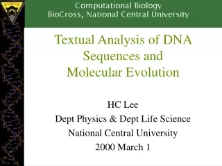
Pairwise alignments in the 1950s b-corticotropin (sheep) Corticotropin A (pig) ala gly glu asp asp glu asp gly ala glu asp glu CYIQNCPLG CYFQNCPRG Oxytocin Vasopressin Early alignments revealed --differences in amino acid sequences between species --differences in amino acids responsible for distinct functions
Pairwise sequence alignment is the most fundamental operation of bioinformatics • • It is used to decide if two proteins (or genes) • are related structurally or functionally • • It is used to identify domains or motifs that • are shared between proteins • It is the basis of BLAST searching (next week) • • It is used in the analysis of genomes
Pairwise alignment: protein sequences can be more informative than DNA • • protein is more informative (20 vs 4 characters); • many amino acids share related biophysical properties • • codons are degenerate: changes in the third position • often do not alter the amino acid that is specified • • protein sequences offer a longer “look-back” time • (relatedness over millions or billions of years) • (note: issue of convergent evolution) • DNA sequences can be translated into protein, • and then used in pairwise alignments
Pairwise alignment: protein sequences can be more informative than DNA • DNA can be translated into six potential proteins 5’ CAT CAA 5’ ATC AAC 5’ TCA ACT 5’ CATCAACTACAACTCCAAAGACACCCTTACACATCAACAAACCTACCCAC 3’ 3’ GTAGTTGATGTTGAGGTTTCTGTGGGAATGTGTAGTTGTTTGGATGGGTG 5’ 5’ GTG GGT 5’ TGG GTA 5’ GGG TAG
Pairwise alignment: protein sequences can be more informative than DNA • Many times, DNA alignments are appropriate • --to confirm the identity of a cDNA • --to study noncoding regions of DNA • --to study DNA polymorphisms • --to study molecular evolution (syn. vs nonsyn) • --example: Neanderthal vs modern human DNA Query: 181 catcaactacaactccaaagacacccttacacccactaggatatcaacaaacctacccac 240 |||||||| |||| |||||| ||||| | ||||||||||||||||||||||||||||||| Sbjct: 189 catcaactgcaaccccaaagccacccct-cacccactaggatatcaacaaacctacccac 247
Definitions Pairwise alignment The process of lining up two or more sequences to achieve maximal levels of identity (and conservation, in the case of amino acid sequences) for the purpose of assessing the degree of similarity and the possibility of homology.
Definitions Homology Similarity attributed to descent from a common ancestor. Identity The extent to which two (nucleotide or amino acid) sequences are invariant. RBP 26 RVKENFDKARFSGTWYAMAKKDPEGLFLQDNIVAEFSVDETGQMSATAKGRVRLLNNWD- 84 +K++ +++ GTW++MA + L + A V T + +L+ W+ glycodelin 23 QTKQDLELPKLAGTWHSMAMA-TNNISLMATLKAPLRVHITSLLPTPEDNLEIVLHRWEN 81
Definitions Conservation Changes at a specific position of an amino acid or (less commonly, DNA) sequence that preserve the physico-chemical properties of the original residue. Similarity The extent to which nucleotide or protein sequences are related. It is based upon identity plus conservation.
Definitions: two types of homology Orthologs Homologous sequences in different species that arose from a common ancestral gene during speciation; may or may not be responsible for a similar function. Paralogs Homologous sequences within a single species that arose by gene duplication.
Pairwise GLOBAL alignment of retinol-binding protein and b-lactoglobulin 1 MKWVWALLLLAAWAAAERDCRVSSFRVKENFDKARFSGTWYAMAKKDPEG 50 RBP . ||| | . |. . . | : .||||.:| : 1 ...MKCLLLALALTCGAQALIVT..QTMKGLDIQKVAGTWYSLAMAASD. 44 lactoglobulin 51 LFLQDNIVAEFSVDETGQMSATAKGRVR.LLNNWD..VCADMVGTFTDTE 97 RBP : | | | | :: | .| . || |: || |. 45 ISLLDAQSAPLRV.YVEELKPTPEGDLEILLQKWENGECAQKKIIAEKTK 93 lactoglobulin 98 DPAKFKMKYWGVASFLQKGNDDHWIVDTDYDTYAV...........QYSC 136 RBP || ||. | :.|||| | . .| 94 IPAVFKIDALNENKVL........VLDTDYKKYLLFCMENSAEPEQSLAC 135 lactoglobulin 137 RLLNLDGTCADSYSFVFSRDPNGLPPEAQKIVRQRQ.EELCLARQYRLIV 185 RBP . | | | : || . | || | 136 QCLVRTPEVDDEALEKFDKALKALPMHIRLSFNPTQLEEQCHI....... 178 lactoglobulin 25% identity; 32% similarity
RBP and b-lactoglobulin are homologous proteins that share related three-dimensional structures b-lactoglobulin (P02754) retinol-binding protein (NP_006735)
Gaps • Positions at which a letter is paired with a null are called gaps. • Gap scores are typically negative. • Since a single mutational event may cause the insertion or deletion of more than one residue, the presence of a gap is ascribed more significance than the length of the gap. • In BLAST, it is rarely necessary to change gap values from the default.
Should distantly related species have more gaps than closely related species (or genes)? What about their relationship in regards to sequence identity?
There are 3 Principal Methods of Pair-wise Sequence Alignment • Dot Matrix Analysis (e.g. Dotlet, Dotter, Dottup) • Dynamic Programming (DP) algorithm • Word or k-tuple methods (e.g. FASTA & BLAST)
General approach to pairwise alignment • Choose two sequences • Select an algorithm that generates a score • Allow gaps (insertions, deletions) • Score reflects degree of similarity • Alignments can be global or local • Estimate probability that the alignment • occurred by chance
Calculation of an alignment score http://www.ncbi.nlm.nih.gov/Education/BLASTinfo/Alignment_Scores2.html
Scoring Matrices take into account: Conservation – acceptable substitutions while not changing function of protein (charge, size, hydrophobicity) Frequency – reflect how often particular residues occur among entire collection of proteins (rare residues given more weight) Evolution – different scoring matrices are designed to either detect closely related or more distantly related proteins
Two kinds of sequence alignment: global and local global alignment algorithm --Needleman and Wunsch (1970) local alignment algorithm --Smith and Waterman (1981)
Global alignment versus local alignment Global alignment (Needleman-Wunsch) extends from one end of each sequence to the other Local alignment finds optimally matching regions within two sequences (“subsequences”) Local alignment is almost always used for database searches such as BLAST. It is useful to find domains (or limited regions of homology) within sequences Smith and Waterman (1981) solved the problem of performing optimal local sequence alignment. Other methods (BLAST, FASTA) are faster but less thorough.
Global alignment with the algorithm of Needleman and Wunsch (1970) • • Two sequences can be compared in a matrix • along x- and y-axes. • • If they are identical, a path along a diagonal • can be drawn • • Find the optimal subpaths, and add them up to achieve • the best score. This involves • --adding gaps when needed • --allowing for conservative substitutions • --choosing a scoring system (simple or complicated) • N-W is guaranteed to find optimal alignment(s)
Three steps to global alignment with the Needleman-Wunsch algorithm [1] set up a matrix [2] score the matrix [3] identify the optimal alignment(s)
Four possible outcomes in aligning two sequences 1 2 [1] identity (stay along a diagonal) [2] mismatch (stay along a diagonal) [3] gap in one sequence (move vertically!) [4] gap in the other sequence (move horizontally!)
How the Smith-Waterman algorithm works Set up a matrix between two proteins (size m+1, n+1) No values in the scoring matrix can be negative! S > 0 The score in each cell is the maximum of four values: [1] s(i-1, j-1) + the new score at [i,j] (a match or mismatch) [2] s(i,j-1) – gap penalty [3] s(i-1,j) – gap penalty [4] zero
Rapid, heuristic versions of Smith-Waterman: FASTA and BLAST Smith-Waterman is very rigorous and it is guaranteed to find an optimal alignment. But Smith-Waterman is slow. It requires computer space and time proportional to the product of the two sequences being aligned (or the product of a query against an entire database). Gotoh (1982) and Myers and Miller (1988) improved the algorithms so both global and local alignment require less time and space. FASTA and BLAST provide rapid alternatives to S-W
How FASTA works [1] A “lookup table” is created. It consists of short stretches of amino acids (e.g. k=3 for a protein search). The length of a stretch is called a k-tuple. The FASTA algorithm finds the ten highest scoring segments that align to the query. [2] These ten aligned regions are re-scored with a PAM or BLOSUM matrix. [3] High-scoring segments are joined. [4] Needleman-Wunsch or Smith-Waterman is then performed.
Pairwise alignment: BLAST 2 sequences • Go to http://www.ncbi.nlm.nih.gov/BLAST • Choose BLAST 2 sequences • In the program, [1] choose blastp or blastn [2] paste in your accession numbers (or use FASTA format) [3] select optional parameters --3 BLOSUM and 3 PAM matrices --gap creation and extension penalties --filtering --word size [4] click “align”
homologous sequences non-homologous sequences Sequences reported as related True positives False positives Sequences reported as unrelated False negatives True negatives Sensitivity: ability to find true positives Specificity: ability to minimize false positives
Substitution Matrix A substitution matrix contains values proportional to the probability that amino acid i mutates into amino acid j for all pairs of amino acids. Substitution matrices are constructed by assembling a large and diverse sample of verified pairwise alignments (or multiple sequence alignments) of amino acids. Substitution matrices should reflect the true probabilities of mutations occurring through a period of evolution. The two major types of substitution matrices are PAM and BLOSUM.
Normalized frequencies of amino acids: variations in frequency of occurrence Gly 8.9% Arg 4.1% Ala 8.7% Asn 4.0% Leu 8.5% Phe 4.0% Lys 8.1% Gln 3.8% Ser 7.0% Ile 3.7% Val 6.5% His 3.4% Thr 5.8% Cys 3.3% Pro 5.1% Tyr 3.0% Glu 5.0% Met 1.5% Asp 4.7% Trp 1.0% blue=6 codons; red=1 codon; note: should be 5% for each if equally distributed
Dayhoff et al. examined multiple sequence alignments (e.g. glyceraldehyde 3-phosphate dehydrogenases) to generate tables of accepted point mutations fly GAKKVIISAP SAD.APM..F VCGVNLDAYK PDMKVVSNAS CTTNCLAPLA human GAKRVIISAP SAD.APM..F VMGVNHEKYD NSLKIISNAS CTTNCLAPLA plant GAKKVIISAP SAD.APM..F VVGVNEHTYQ PNMDIVSNAS CTTNCLAPLA bacterium GAKKVVMTGP SKDNTPM..F VKGANFDKY. AGQDIVSNAS CTTNCLAPLA yeast GAKKVVITAP SS.TAPM..F VMGVNEEKYT SDLKIVSNAS CTTNCLAPLA archaeon GADKVLISAP PKGDEPVKQL VYGVNHDEYD GE.DVVSNAS CTTNSITPVA fly KVINDNFEIV EGLMTTVHAT TATQKTVDGP SGKLWRDGRG AAQNIIPAST human KVIHDNFGIV EGLMTTVHAI TATQKTVDGP SGKLWRDGRG ALQNIIPAST plant KVVHEEFGIL EGLMTTVHAT TATQKTVDGP SMKDWRGGRG ASQNIIPSST bacterium KVINDNFGII EGLMTTVHAT TATQKTVDGP SHKDWRGGRG ASQNIIPSST yeast KVINDAFGIE EGLMTTVHSL TATQKTVDGP SHKDWRGGRT ASGNIIPSST archaeon KVLDEEFGIN AGQLTTVHAY TGSQNLMDGP NGKP.RRRRA AAENIIPTST fly GAAKAVGKVI PALNGKLTGM AFRVPTPNVS VVDLTVRLGK GASYDEIKAK human GAAKAVGKVI PELNGKLTGM AFRVPTANVS VVDLTCRLEK PAKYDDIKKV plant GAAKAVGKVL PELNGKLTGM AFRVPTSNVS VVDLTCRLEK GASYEDVKAA bacterium GAAKAVGKVL PELNGKLTGM AFRVPTPNVS VVDLTVRLEK AATYEQIKAA yeast GAAKAVGKVL PELQGKLTGM AFRVPTVDVS VVDLTVKLNK ETTYDEIKKV archaeon GAAQAATEVL PELEGKLDGM AIRVPVPNGS ITEFVVDLDD DVTESDVNAA Examined 1572 changes in 71 groups of closely related proteins
Dayhoff’s PAM1 mutation probability matrix Original amino acid Each element of the matrix shows the probability that an amino acid (top) will be replaced by another residue (side) (n=10,000)
Dayhoff’s PAM1 mutation probability matrix (Point-Accepted Mutations) • All the PAM data come from alignments of closely • related proteins (>85% amino acid identity) • PAM matrices are based on global sequence alignments. • The PAM1 is the matrix calculated from comparisons • of sequences with no more than 1% divergence • (all other PAM matrices are extrapolated from PAM1). • For the PAM1 matrix, that interval is 1% amino acid • Divergence; note that the interval is not in units of time.
PAM0 and PAM2000 mutation probability matrices Consider a PAM0 matrix. No amino acids have changed, so the values on the diagonal are 100%. Consider a PAM2000 (nearly infinite) matrix. The values approach the background frequencies of the amino acids (given in Table 3-2).
The PAM250 mutation probability matrix The PAM250 matrix is of particular interest because it corresponds to an evolutionary distance of about 20% amino acid identity (the approximate limit of detection for the comparison of most proteins). Note the loss of information content along the main diagonal, relative to the PAM1 matrix.
PAM250 mutation probability matrix Top: original amino acid Side: replacement amino acid
Why do we go from a mutation probability matrix to a log odds matrix? • We want a scoring matrix so that when we do a pairwise • alignment (or a BLAST search) we know what score to • assign to two aligned amino acid residues. • Logarithms are easier to use for a scoring system. They • allow us to sum the scores of aligned residues (rather • than having to multiply them).
How do we go from a mutation probability matrix to a log odds matrix? • The cells in a log odds matrix consist of an “odds ratio”: • the probability that an alignment is authentic • the probability that the alignment was random • The score S for an alignment of residues a,b is given by: • S(a,b) = 10 log10 (Mab/pb) • As an example, for tryptophan, • S(a,tryptophan) = 10 log10 (0.55/0.010) = 17.4
PAM250 log odds scoring matrix
What do the numbers mean in a log odds matrix? S(a,tryptophan) = 10 log10 (0.55/0.010) = 17.4 A score of +17 for tryptophan means that this alignment is 55 times more likely than a chance alignment of two Trp residues. S(a,b) = 17 Probability of replacement (Mab/pb) = x Then 17.4 = 10 log10 x 1.74 = log10 x 101.74 = x = 55