Download
1 / 31
380 likes | 1.09k Views
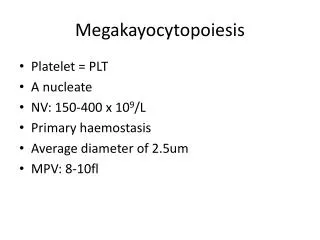
Megakayocytopoiesis. Platelet = PLT A nucleate NV: 150-400 x 10 9 /L Primary haemostasis Average diameter of 2.5um MPV: 8-10fl. Platelets . Arise from megakaryocytes Largest cells in the body (30-50um) Polyploid(chromosomes more than 46) Cluster on extravascular compartment.

E N D
Megakayocytopoiesis • Platelet = PLT • A nucleate • NV: 150-400 x 109/L • Primary haemostasis • Average diameter of 2.5um • MPV: 8-10fl
Platelets • Arise from megakaryocytes • Largest cells in the body (30-50um) • Polyploid(chromosomes more than 46) • Cluster on extravascular compartment
Megakayocyte Progenitor • CFU-GEMM • Thrombopoietin • BFU-Meg • CFU-Meg • LD-CFU-Meg enters TERMINAL DIFFERENTIATION
Megakayocyte Progenitor • Developmental Compartments: • Proliferative Stage • Terminal Differentiation
Megakayocyte Progenitor • Colony Forming Units Megakaryocyte • Resembles a lymphocyte • Diploid(chromosome double No.) • Participates in mitosis • Burst Forming Units Megakaryocyte • Resembles a lymphocyte • Diploid • Participates in mitosis
Megakayocyte Progenitor • Light Density colony forming unit Meg • Low mitotic capacity • Increased nuclear ploidy • Transitional or “Promegakayoblast”
Megakayocyte Progenitor • Terminal Differentiation • Goes through 3 different stages • MKI: Megakaryoblast • MKII: Promegakaryocyte • MKIII: Megakaryocyte
Endoreduplication • Also known as endomitosis • Mitosis without telophase and cytokinesis • Single megarkaryocyte can give rise to 2000 to 4000 platelets • Segmentation of MKII and MKIII reflects endomitosis
Thrombopoiesis • DMS (demarcation system) dilates • Longitudinal bundles of tubules are formed • Proplatelet process extends • Transverse constriction • Proplatelet process pierces through the sinusoidal lining of endothelial cells • Extends into the venous blood • Releases platelet
Megakayocyte Progenitor • Thrombopoietin • 70 KD molecule with 23% homology to EPO • Produced in the liver • Ligand that binds megakaryocytes and platelet membrane receptor protein • Concentration is inversely proportion to the platelet and megakaryocyte mass • Induces stem cell differentiation • In vitro studies shows that it increases the amount of platelet in the bone marrow in healthy subjects
Megakayocyte Progenitor • Cell derived stimulators or megakaryopoiesis: • Interleukin 3: acts in synergy with TPO; induces early differentiation of stem cells • Interleukin 6: acts only in the presence of TPO; enhances endomitosis. Megakaryocyte maturation and platelet release • Interleukin 11: stimulate platelet production in chemotherapy induced thrombocytopenia
Overview Platelet Structure • Platelet Structure: Peripheral Zone • Glycocalyx • Plasma membrane • Lipids • Receptors • SCCS(surface connected canalicular system) • DTS(dense tubular system) • Platelet Structure: Sol-Gel Zone • Cytoskeleton • Microtubules • Microfilaments • Platelet Structure: Organelle Zone • Cytoplasmic Content • PLT Granules • Alpha • Dense • Lysosomes
Overview: Platelet Structure • Anucleatecytoplasmic pieces shed from megakaryocyte • Metabolically active • Phospholipid source for clot formation • Release chemicals that mediate physiological response to tissue damage leading to hemorrhage, thrombosis and tissue repair • House receptors that facilitate PLT activation and PLT participation in primary and secondary hemostasis • 1/3rd PLTs released from BM are sequestered by spleen • PB Ref range: 150 – 450 x 109/L Graphic accessed at URL http://hsc.virginia.edu/medicine/clinical/pathology/educ/innes/text/nh/platelets.html & http://www.med-ed.virginia.edu/courses/path/innes/images/nhjpeg/nh%20megakaryocyte%20x50a.jpeg
Platelet Structure - Peripheral Zone • Glycocalyx • Plasma Membrane - Bilayered • Lipids • Receptors • Surface-connected canalicular system (SCCS) • Dense tubular System (DTS) Graphics accessed URL http://evolvels.elsevier.com/section/default.asp?id=1138_ccalvo7_0001, 2008.
Glycocalyx • Amorphous exterior coat • Negatively charged • Site of PLT functional environmen • PLT antigenicity • Adhesion receptors Electron micrographs from a resting platelet (x10,000), or from an activated platelet showing pseudopodia emission (x5,000). (Pictures have been kindly provided by Dr. J. White). Graphic accessed http://www.platelet-research.org/2/morph_rest.htm, 2009.
PLT Plasma Membrane: Lipids • Plasma layer • Cytoplasmic layer • Sources of arachidonic acid • PLT activation Graphic accessed URL http://en.wikipedia.org/wiki/File:Eicosanoid_synthesis.svg 2008.
PLT Plasma Membrane: Receptors • Mediate adhesion & aggregation • Facilitate secretion Graphic accessed URL http://www.platelets.se/resource/Platelet%20function.jpg, 2009.
M&M graphic accessed/adapted URL http://www.tu-pc.com/fondos/media/2452.jpg. 2009. PLT Plasma Membrane: SCCS • Tubular invagination of membrane • glycocalyx • Transport of substances released from PLT granules to exterior • Adsorption/storage of hemostatic proteins White, JG and Clawson, CC. The surface-connected canalicular system of blood platelets: a fenestrated membrane system (1980). Am Journal of Pathology, 101, 353-364, and Graphic accessed URL http://www.chelationtherapyonline.com/GarryGordon/images/Copy%20(3)%20of%20Slide1.GIF , 2009.
Dense Tubular System (DTS) • Endoplasmic reticulum remnant • From megakaryocyte • Calcium storage/release • Prostaglandin & Thromboxane synthesis Histochemistry (1982) 76:189-196 , Am J Pathol. 1976 May; 83(2): 283–298 & Thromb Haemost. 1978 Oct 31;40(2):224-31 Graphic accessed URL http://www.platelet-research.org/2/morph_rest.htm, 2009.
Platelet Structure: Sol-Gel Zone • PLT Cytoskeleton • Microtubules • Tubulin • Microfilaments • Actin/Myosin • Contractile protein: PLT shape-change Graphic accessed URL http://health.upenn.edu/News/News_Releases/platelet.jpg, 2009.
Platelet Structure: Organelle-Zone • Cytoplasmic content • Anucleate • Few mitochondria • Granules • Alpha • Dense • Lysosomes Graphic accessed URL http://www.platelet-research.org/2/morph_rest.htm, 2009.
PLT Granules’ Content Primary hemostasis, Secondary hemostasis
Hemostasis Def. mechanism by which blood is kept inside the blood vesseles(no hemorrhage) in fluid state(no thrombosis.
Overview: Platelet Function • PRIMARY HEMOSTASIS • Form platelet plug • SECONDARYHEMOSTASIS • Reaction surface for coagulation Graphic accessed at URL http://www.medicine.mcgill.ca/physio/209A/Blood/blood6a.htm, 2007.
PLT Function: Vascular Integrity • Inner layer- monolayer of endothelia • Middle layer – smooth muscle, connective tissue • Vasoconstriction • Outer layer– fibroblasts, collagen, mast cells • Tissue factor source • The main response of blood vessels in order to hemostasis is a vasoconstriction Graphic accessed at URL http://cellapplications.com/images/humanimages/HCtAEC2115-s2.jpg, 9/12/08.
Platelet Plug Formation:Adhesion • Platelets bind to exposed adhesive subendothelial connective tissue • Collagen • vWF • Fibronectin • Mechanism components • vWF: links PLT to endothelial binding site • PLT receptor GPIb IX • Collagen fibers • Actin contracts & pseudopods form • REVERSIBLE MECHANISM • Facilitates activation M&M graphic accessed http://www.dicksons.com.au/images/body/products/prod_mm_guys.gif, 2009.
M&M graphic accessed URL http://www.jauntyquills.com/pics/WebPics/M&Mgroup.jpg, 2009. Platelet Plug Formation: Aggregation • Platelet-Platelet interaction • Mechanism components • ATP • Ionized calcium • Fibrinogen • PLT receptor GPIIb/IIIa • Initial aggregation – REVERSIBLE MECHANISM • Secondary aggregation - IRREVERSIBLE = white clot, a platelet plug formed. Graphic accessed URL http://www.medscape.com/content/2003/00/45/62/456257/art-jic456257.fig1.jpg, 2008.
Platelet Plug Formation: Secretion • Discharge of granules’ contents • Markers of PLT activation • PF4 • PDGF • Thromboglobulin • Promote & Amplify PLT activities • Primary hemostasis • Secondary hemostasis Graphic accessed http://www.uptodate.com/online/content/images/hema_pix/Platelet_interactions.jpg, 2009.
Summary: PLT Form and Function • Platelets are derived from BM megakaryocytes. • Circulating platelets are anucleate, but have a distinctive anatomy and physiology that enables them to play important roles in primary and secondary hemostasis. • The PLT membrane houses numerous receptors that control PLT activation. • PLT function can be inhibited by drugs which target key PLT activation enzymes and receptors. • PLTs adhere to surfaces, aggregate with each other, and secrete their granules’ contents in order to plug damaged areas in endothelia, modulate coagulation, and aid tissue repair.