Download
1 / 18
180 likes | 328 Views
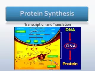
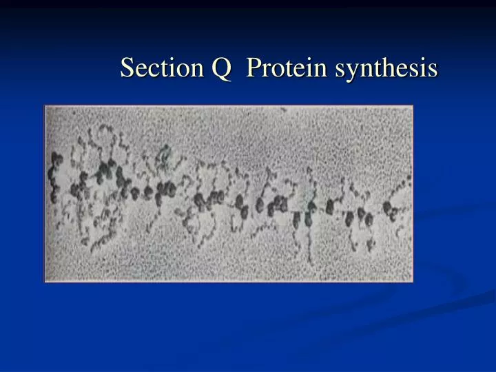
Section Q Protein synthesis. content. Q1 ASPECTS OF PROTEIN SYNTHESIS Q2 MECHANISM OF PROTEIN SYNTHESIS Q3 INITIATION IN EUKARYOTES Q4 TRANSLATIONAL CONTROL AND POST-TRANSLATIONAL EVENTS. Q1 ASPECTS OF PROTEIN SYNTHESIS.

E N D
content • Q1 ASPECTS OF PROTEIN SYNTHESIS • Q2 MECHANISM OF PROTEIN SYNTHESIS • Q3 INITIATION IN EUKARYOTES • Q4 TRANSLATIONAL CONTROL AND POST-TRANSLATIONAL EVENTS
Q1 ASPECTS OF PROTEIN SYNTHESIS • Codon-anticodon interactionIn the cleft of the ribosome, an antiparallel formation of three pairs occurs between the codon on the mRNA and the anticodon on the tRNA. If the 5’ anticodon base is modified, the tRNA can usually interact with more than one codon. • Wobble The wobble hypothesis decribes the nonstandard base pairs that can form between modified 5’-acticodon bases and 3’-codon bases. When the wobble nucleoside is inosine, the tRNA can base-pair with three codon-those ending in A, C or U.
Ribosome binding site The ribosome binding site is a sequence just upstream of the initiation codon in prokaryotic mRNA which base-pairs with a complementary sequence near the 3’-end of the 16S rRNA to position the ribosome for initiation of protein synthesis. It is also known as the Shine-Dalgarno sequence after its discoverers. • Polysome Polyribosomes(polysomes) form on an mRNA when successive ribosomes attach, begin translating and move along the mRNA. A polysome is a complex of multiple ribosomes in various stages of translation on one mRNA molecule. • Initiator tRNA A special tRNA(initiator tRNA), recognizingh the AUG start codon, is used to initiate protein synthesis in both prokaryotes and eukaryotes. In prokaryotes, the initiator tRNA is first charged with methionine by methionyl-tRNA synthetase. The methionine residue is then converted to N-formylmethionine by transformylase. In eukaryotes, the methionine on the initiator tRNA is not modified. There are strucutral differences between the E.coli initiator tRNA and the tRNA that insects internal Met residues.
Fig. 2. The E. coli methionine-tRNAs. (a) The initiator tRNA fMet-tRNAfMet; (b) the methionyl-tRNA Met-tRNAMet
Q2 MECHANISM OF PROTEIN SYNTHESIS • OverviewThere are three stages of protein synthesis: initiation—the assembly of a ribosome on an mRNA; elongation—repeated cycles of amino acid delivery, peptide bond formation movement along the mRNA(translation); termination—the relase of the polypeptide chain. • InitiationIn prokaryotes, initiation requires the large and small ribosome subunits, the mRNA, the initiator tRNA, three initiation factor(IFs) and GTP. IF1 and IF3 bind to the 30S subunit and prevent the large subunit binding. IF2+GTP can then bind and will help the initiator tRNA to bind later. This small subunit complex can now attach to an mRNA via its ribosome-binding site. The initiator tRNA can then base-pair with the AUG initiation codon which releases IF3 thus creating the 30S initiation complex. The large subunit then binds, displacing IF1 and IF2+GTP, giving the 70S initiation comple which is the fully assembled ribosome at the correct position on the mRNA.
Elongation Elongation involves the three factors(Efs), EF-Tu, EF-Ts and EF -G, GTP, charged tRNA and the 70S initiation complex(or its equivalent). It takes place in three steps. A charged tRNA is delivered as a complex with EF-Tu and GTP. The GTP is hydrolyzed and EF-Tu.GTP is released which can be re-used with the help of EF-Ts and GTP(via the EF-Tu-EF-Ts exchange cycle). Peptidyl transferase makes a peptide bond by joining the two adjacent amino acid withou t the input of more energy. Translaocase(EF-G), with energy from GTP, move the ribosome one codon along the mRNA, ejecting the uncharged tRNA and transferring the growing peptide chain to the P-site. • Termination Release factors(RF1 or RF2) recognize the stop codon and, helped by RF3, make peptidyl transferase join the polypeptide chain to a water molecule, thus releasing it. Ribosome release factor helps to dissociate the ribosome subunit from the mRNA.
Fig. 1. Initiation of protein synthesis in the prokaryote E. coli.
Q3 INITIATION IN EUKARYOTES • OverviewMost of the differences in the mechanism of protein synthesis between prokaryotes and eukaryotes occur in the initiation stage; however, eukaryotes have just one release factor(eRF). The eukaryotic initiator tRNA does not become N-formaylated as in prokaryotes. • ScanningThe eukaryotic 40S ribosome subunit complex binds to the 5’-cap region of the mRNA complex and moves along it looking(scanning) for an AUG start codon. It is not always the first AUG, as it must have appropriate sequences around it.
InitiationThis is the major point of difference between prokaryotic and eukaryotic protein synthesis, there being at least nine eIF involved. Functionally, these factors can be grouped. They either bind to the ribosome subunit or to the mRNA, deliver the initiator tRNA or didplace other other factors. In contrast to the events in prokaryotes, initiation involves the initiator tRNA binding to the 40S subunit before it can bind to the mRNA. Phosphorylation of eIF2, which delivers the initiator tRNA, is an important control point. • ElongationThis stage of protein synthesis is essentially identical to that described for prokaryotes. The factors EF-Tu, EF-Ts and EF-G have direct eukaryotic equivalents called eEF1α, eEF1βγ and eEF2 respectively, which carry out the same roles. • TerminationEukaryotes use only one release factor(eRF), which requires GTP, for termination of protein synthesis. It can recognize all three codons.
Tble 1.Comparison of peoyein synthesis factors in prokaryotes and eukaryotes
Q4 TRANSLATIONAL CONTROL AND POST-TRANSLATIONAL EVENTS • Translational controlIn prokaryotes, the level of translation of different cistrons canbe affected by: (i) the binding of short antisense molecules, (ii) the relative stability to nucleases of part of the polycistronic mRNA, and (iii) the binding of proteins that prevent ribosome access. In eukaryotes, protein binding can also mask the mRNA and prevent translation, and repeats of the sequence 5’-AUUUA-3’ can make the mRNA unstable and less frequently translated. • PolyproteinsA single translation product that is cleaved to generate two more separate proteins is called a polyprotein. Many viruss produce polyproteins.
Protein targetingCertain short peptide sequences in proteins determine the cellular location of the protein, such as nucleus, mitochondrion or chloroplast. The signal sequence of secreted proteins cause the translating ribosome to bind factors that make the ribosome dock with a membrane and transfer the protein through the membrane as it is synthesized. Usually the signal sequence is then cleaved off by signal peptidase. • Protein modificationThe most common alterations to nascent polypeptides are those of cleavage and chemical modification. Cleavage occurs to remove signal peptides, to release mature fragments from polyproteins, to remove internal peptides as well as trimming both N- and C-termini. There are many chemical modifications that can take place on all but six of the amino acid side chains. Often phosphorylation controls the activity of protein. • Protein degradationDamaged, modification or inherently unstable proteins are marked for degradation by having multiole molecules of ubiquitin covalently attached. The ubiquitinylated protein is then degraded by a 26S protease complex.