Download
1 / 11
110 likes | 206 Views
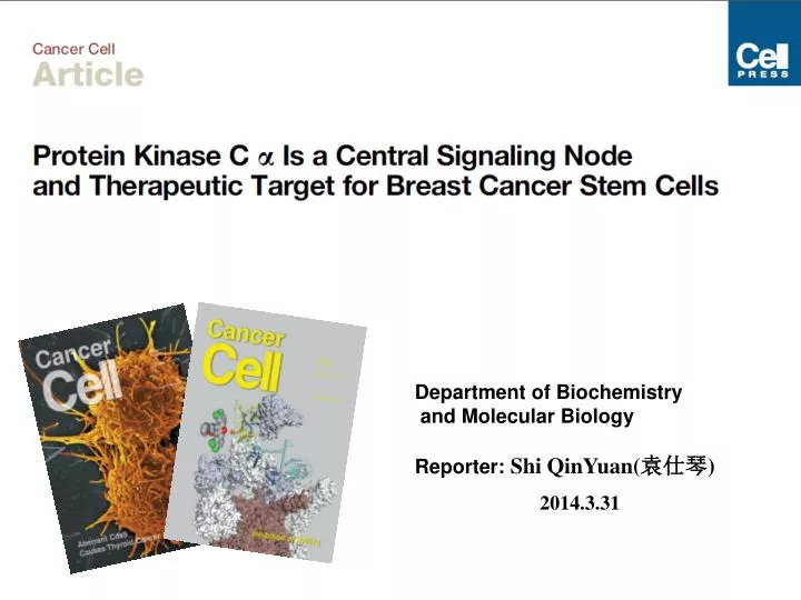
Department of Biochemistry and Molecular Biology Reporter: Shi QinYuan( 袁仕琴 ) 2014.3.31. Introduction:. What can we do for the cancer stem cell(CSCs ) ?. Significance:. Design:. To discover key regulatory genes unique to the mesenchymal state Whose expression is elevated in CSCs.

E N D
Department of Biochemistry and Molecular Biology Reporter: Shi QinYuan(袁仕琴) 2014.3.31
Introduction: What can we do for the cancer stem cell(CSCs ) ?
Design: To discover key regulatory genes unique to the mesenchymal state Whose expression is elevated in CSCs. Downsteam signals of PKCa__FRA1 Upsteam signals of PKCa__PDGFR Therapeutic effects of PKCa inhibition Identification of kinases expressed differentially in EMT-Induced cells Build a EMT model Cancer Cell PKCa Signaling Network in Breast Cancer Stem Cells
Result:1 Identification of kinases expressed differentially in EMT Induced cells PKCa HMLE(green) , HMLE-twist,snail,slug(red) The inhibitors targeting PKCa, CLK1, CDK6, and JAK1 also appeared to deplete NAMEC-Tom cells preferentially. Using microarray gene expression analyses: A group of kinase-encoding genes was overexpressed at least 2-fold in HMLE-Twist, HMLE-Snail, and HMLE-Slug cells relative to the HMLE population Cancer Cell PKCa Signaling Network in Breast Cancer Stem Cells
Result:3 Therapeutic effects of PKCa inhibition These observations confirmed the greater dependence on PKCa-regulated signaling networks in cells that have passed through an EMT program. Cancer Cell PKCa Signaling Network in Breast Cancer Stem Cells
Result:4 Upsteam signals of PKCa__PDGFR We monitored the activity of PDGFR in the mesenchymal cell populations Cancer Cell PKCa Signaling Network in Breast Cancer Stem Cells
Result:5 Downsteam signals of PKCa__FRA1 Cancer Cell PKCa Signaling Network in Breast Cancer Stem Cells
Conclusion: (1) The EMT program becomes activated during malignant progression and can enrich for cancer stem cells (CSCs). (2) The inhibition of protein kinase C a (PKCa) specifically targets CSCs but has little effect on non-CSCs. (3) The formation of CSCs from non-stem cells involves a shift from EGFR to PDGFR signaling and results in the PKCa-dependent activation of FRA1. (3) We identified an AP-1molecular switch in which c-FOS and FRA1 are preferentially utilized in non-CSCs and CSCs, respectively. (4) PKCa and FRA1 expression is associated with the aggressive triple-negative breast cancers, and the depletion of FRA1 results in a mesenchymal-epithelial transition. (5) Hence, identifying molecular features that shift between cell states can be exploited to target signaling components critical to CSCs. Cancer Cell PKCa Signaling Network in Breast Cancer Stem Cells
References: 1, Thiery, J.P., Acloque, H., Huang, R.Y., and Nieto, M.A. (2009).Epithelialmesenchymal transitions in development and disease. Cell 139, 871–890. 2, Morel, A.P., Lie` vre, M., Thomas, C., Hinkal, G., Ansieau, S., and Puisieux, A. (2008). Generation of breast cancer stem cells through epithelial-mesenchymal transition. PLoS ONE 3, e2888. 3, Kang, J.H., Toita, R., Kim, C.W., and Katayama, Y. (2012). Protein kinase C (PKC) isozyme- specific substrates and their design. Biotechnol Adv. 30,1662–1672. 4, Stinson, S., Lackner, M.R., Adai, A.T., Yu, N., Kim, H.J., O’Brien, C., Spoerke, J., Jhunjhunwala, S., Boyd, Z., Januario, T., et al. (2011). TRPS1 targeting by miR-221/222 promotes the epithelial-to-mesenchymal transition in breastcancer. Sci. Signal. 4, ra41. 5, Nieto, M.A. (2011). The ins and outs of the epithelial to mesenchymal transition in health and disease. Annu. Rev. Cell Dev. Biol. 27, 347–376. 6, Vogelstien B, Kinzler K W. The multistep nature of cancer. Trends Genet,1993,9:138 7, HamburgerAW,Salmon SE.Primary bioassay of human tumor stem cells.Science,1977, 97(4302):461-463.