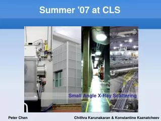
Summer '07 at CLS
140 likes | 265 Views
This project focuses on employing Small Angle X-Ray Scattering (SAXS) as an alternative imaging technique to Scanning Transmission X-Ray Microscopy (STXM) for enhancing structural analysis of samples. We aim to develop software that optimizes camera setup and controls various experimental variables such as exposure settings and sample orientation. The task includes creating visual representations from diffraction patterns and assessing the stability of received data through phase reconstruction. Our goal is to provide insightful imaging capabilities for smaller samples and improve measurement accuracy.

Summer '07 at CLS
E N D
Presentation Transcript
Small Angle X-Ray Scattering Summer '07 at CLS Peter Chen Chithra Karunakaran & Konstantine Kaznatcheev
to provide an alternative imaging technique to STXM : to understand structural information of sample Purpose (Feser/Jacobsen) (Miao, Charalambous, Kirz, Sayre, Nature 400, 342 (1999))
Reconstruction diffraction pattern hologram image SEM image of letters, fabricated by gold dots, (100 nm in diameter each); diffraction pattern (middle) and reconstructed image (Miao, Charalambous, Kirz, Sayre, Nature 400, 342 (1999)). Only intensity is obtained, phase information is lost.
Purpose Mie polar diagrams • to image smaller samples • to test stability and integrity of the optics and the microscope small x=pa/l large
Tasks: create software determine camera setup in the STXM experimentation take data with respect to different variables orientation geometry nature of samples Tasks Andor Vacuum supported CCD. 512 x 512 12.3 x 12.3 mm
Software Visual Basic .NET 2003 Requirements basic: single imaging, video, exposure settings, etc. long exposure aXis readable data and header output beamline read and control Software
Mechanics • Implementation • external placement
SAXS camera OSA sample 1st order beam detector focus zone plate incident beam scattered wave
Scans on the Mesh x x x Exposure: 100 secEnergy: 710 eV
Change in Focus • Defocusing of beam by 10 um by 30 um
Change in Energy Energy: 709 eV Energy: 705 eV Energy: 703 eV
Change in Slit Size 50 um x 50 um slit size 20 um x 20 um slit size 100 um x 100 um slit size 10 um x 10 um slit size
Consistency • Iron Particles Exposure: 100 secEnergy: 710 eV Background Corrected 10 nm particle.