Download
1 / 42
530 likes | 1.04k Views
Monitoring During Thoracic Anesthesia. Outline. Introduction Physiological Aspects Monitoring Requirements. Introduction. Thoracic anesthesia is challenging. Patient. Procedure. Physiological Aspects. Principles of Ventilation and Perfusion.

E N D
Outline • Introduction • Physiological Aspects • Monitoring Requirements
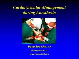
Thoracic anesthesia is challenging Patient Procedure
Principles of Ventilation and Perfusion • “V" - ventilation - the air which reaches the lungs • "Q" - perfusion - the blood which reaches the lungs • Normal V is 4 L of air per minute. • Normal Q is 5L of blood per minute. • So Normal V/Q ratio is 4/5 or 0.8. • When the V/Q is higher than 0.8, it means ventilation exceeds perfusion. • When the V/Q is < 0.8, there is a VQ mismatch caused by poor ventilation
Principles of Ventilation and Perfusion • An area with no ventilation (and thus a V/Q of zero) is termed "shunt." • An area with no perfusion (and thus a V/Q of infinity) is termed “dead space”
Lung Compliance • A change in volume divided by a change in transpulmonary pressure. • (CL = ΔV / ΔPL) • A typical value of compliance is 200 ml/cm H20
Lateral Decubitus Position Optimal access for most operations Alter the normal pulmonary ventilation/perfusion relationships accentuated by Induction of anesthesia Initiation of mech.ventilation Opening the chest Surgical retractions
Principles of Ventilation and Perfusion Perfusion Ventilation Pulmonary blood flow distribution relative to the alveolar pressure
Lateral Decubitus Position The dependent lung is better Ventilated than the Nondependent lung, ˙V/˙ Q still is well matched. Patient awake spontaneously breathing
One Lung Ventilation • The principle physiologic change of OLVis the redistribution of lung perfusion between the ventilated (dependent) and blocked (nondependent) lung • Many factors contribute to the lung perfusion, the major determinants of them are hypoxic pulmonary vasoconstriction, HPV and gravity.
Hypoxic pulmonary vasoconstriction HPV is a widely conserved, homeostatic, vasomotor response of precapillary smooth muscle in the PAs to alveolar hypoxia. HPV mediates ˙V/˙Q matching and, by reducing shunt fraction, optimizes systemic pO2.
One Lung Ventilation Reduces the surface area available for gas exchange Reduced arterial oxygen tension Maintaining oxygenation and elimination of carbon dioxide is the greatest challenge
Monitoring Use of Monitoring to Detect and Diagnose Intraoperative Events • Respiration • Oxygenation • Ventilation • Cardiovascular function
Cardiovascular function • Electrocardiography Arrhythmia, ischemia • Intraarterial catheter Hypotension or hypertension Arterial compression
Cardiovascular function • Pulmonary artery catheter Pulmonary hypertension, filling pressures, assess cardiac performance • SvO2 Adequacy of cardiac output
Cardiovascular function • Transesophageal Echocardiography Ischemia, volume status, right ventricular dysfunction
Tiered Monitoring System of Thoracic Surgery Failure to check the equipment properly before induction of anesthesia is responsible for 22% of the critical incidents that occur during anesthesia
Tiered Monitoring System of Thoracic Surgery Tier I Healthy patients no special intraopertive conditions Sick patients special intraopertive conditions Patient Procedure
Tiered Monitoring System of Thoracic Surgery Tier II Healthy patients no special intraopertive conditions Sick patients special intraopertive conditions Patient Procedure
Tiered Monitoring System of Thoracic Surgery Tier II Healthy patients no special intraopertive conditions Sick patients special intraopertive conditions Patient Procedure
Spirometry • Spirometry is a non-invasive monitor device which measures volume, pressure and flow in the airway. • These measurements may be used to construct : • a pressure-volume curve (PV) and • a flow-volume curve (FV). • The constructed curves will give important information about the peri-operative respiratory function.
Tiered Monitoring System of Thoracic Surgery Tier III Healthy patients no special intraopertive conditions Sick patients special intraopertive conditions Patient Procedure
Special considerations for PA catheter Measured values: • CVP: 1-6 mm Hg (reflects right atrial pressure). • PAP: Systolic 15-30mm Hg, Diastolic 6-12mm Hg. • PCWP: 6 - 12mm Hg. Estimates left atrial heart pressure and left ventricular end diastolic pressure. • CO: 3.5 - 7.5 L/min • Sv02: (70 - 75%). Drawn from the end of the pulmonary artery catheter. Used to calculate how well oxygen is extracted by the tissues.
Special considerations for PA catheter • the LDP is important with regard to pulmonary artery catheter monitoring in three situations. • The catheter is in the nondependent collapsed lung, the measured cardiac output and mixed venous blood (pvo2) may be decreased. • When the nondependent lung is ventilated with PEEP and the catheter is in the nondependent lung, Ppaw may not equal Pla. • When the catheter is in the dependent lung, Ppaw will be a faithful index of Pla, even if PEEP is used
Message to Take Home Monitors are useful adjuncts, But they alone cannot replace Careful observation by Anaesthesiologist.