BACKGROUND
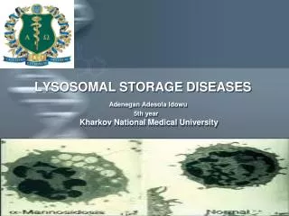
LYSOSOMAL STORAGE DISEASES Adenegan Adesola Idowu 5th year Kharkov National Medical University. BACKGROUND. Lysosomes are subcellular organelles responsible for the physiologic

BACKGROUND
E N D
Presentation Transcript
LYSOSOMAL STORAGE DISEASESAdenegan Adesola Idowu 5th yearKharkov National Medical University
BACKGROUND Lysosomes are subcellular organelles responsible for the physiologic turnover of cell constituents. Lysosomal storage diseases describe a heterogeneous group of dozens of rare inherited disorders characterized by the accumulation of undigested or partially digested macromolecules, which ultimately results in cellular dysfunction and clinical abnormalities. Several studies have explored different methods of treatment, but only are promising and safe even though they are very expensive.
PURPOSE The aim of this work is to describe a few of the various lysosomal storage disorder.
Introduction What are LSDs? • This are a group of approximately 50 rare inherited metabolic disorders that result from defects in lysosomal function. Lysosomal storage diseases result when the lysosome – a specific organelle in the body's cells – malfunctions. • It is commonly referred to as the cell’s recycling center when the lysosome doesn’t function normally, excess products destined for breakdown and recycling are stored in the cell. • Because there are numerous specific deficiencies, storage diseases are usually grouped biochemically by the accumulated metabolite
Lysosomes are membrane-bound organelles that function as the "stomachs" of eukaryotic cells . They are surrounded by a single membrane, have an acidic interior pH level of about 5 maintained by a proton pump, and carry a high content of digestive enzymes. All require an acidic environment to function properly and are called acid hydrolases. Extracellular materials to be degraded in the lysosome are brought into the cell by either pinocytosis or phagocytosis. Intracellular materials, such as old organelles, are brought into a lysosome by a process called autophagy. Basic cell biology
Classification of LSDs I - Defective metabolism of glycosaminoglycans " the mucopolysaccharidoses" MPS I (Hurler, Hurler-Scheie, Scheie) MPS II (Hunter) MPS III (San filipo Types A,B,C and D) MPS IV (Morquio type A and B) MPS VI (Maroteaux-Lamy) MPS VII (Sly) MPS IX (Hyaluronidase deficiency) Multiple Sulfatase deficiency
II - Defective degradation of glycan portion of glycoproteins Aspartylglucosaminuria Fucosidosis, type I and II Mannosidosis Sialidosis, type I and II III - Defective degradation of glycogen Pompe disease
IV - Defective degradation of sphingolipid components Acid sphingomyelinase deficiency (Niemann-Pick A & B) Fabry disease Farber disease Gaucher disease, type I, II and III GM1 gangliosidosis, type I, II and III GM2 gangliosidosis (Tay-Sachs type I, II, III and Sandhoff Krabbe disease Metachromatic leukodystrophy, type I, II and III
V - Defective degradation of polypeptides Pycnodysostosis VI - Defective degradation or transport of cholesterol, cholesterol esters, or other complex lipids Neuronal ceroid lipofuscinosis, type I, II, III and IV
VII - Multiple deficiencies of lysosomal enzymes Galactosialidosis Mucolipidosis, type II and III VIII - Transport and trafficking defects Cystinosis Danon disease Mucolipidosis type IV Niemann-Pick type C Infantile sialic acid storage disease Salla disease
Tay-Sachs Disease and Sandhoff's Disease Tay-Sachs disease • Deficiency of hexosaminidase A results in accumulation of GM2 in the brain.Autosomal recessive; the most common mutations are carried by 1/27 normal adults of Eastern European (Ashkenazi) Jewish origin • Diagnosis is clinical and can be confirmed by enzyme assay • In the absence of effective treatment, management is focused on screening adults of childbearing age in high-risk populations to identify carriers (by way of enzyme activity and mutation testing) combined with genetic counseling.
Gaucher's Disease Gaucher's disease is a sphingolipidosis resulting from glucocerebrosidase deficiency, causing deposition of glucocerebroside and related compounds Diagnosis is by enzyme analysis of WBCs. Gaucher's cells—lipid-laden tissue macrophages in the liver, spleen, lymph nodes, or bone marrow that have a wrinkled tissue-paper appearance—are diagnostic. Enzyme replacement with placental or recombinant glucocerebrosidase is effective in types I and III; there is no treatment for type II.
Mucopolysaccharidoses (MPS) MPS are inherited deficiencies of enzymes involved in glycosaminoglycan breakdown. Glycosaminoglycans (previously termed mucopolysaccharides) are polysaccharides abundant on cell surfaces and in extracellular matrix and structures. Enzyme deficiencies cause extensive bone, soft tissue, and CNS changes. manifestations include coarse facial features, neurodevelopmental delays and regression, joint contractures, organomegaly Diagnosis is confirmed by enzyme analysis of cultured fibroblasts (prenatal) or peripheral WBCs (postnatal). Treatment of MPS type I (Hurler's disease) is enzyme replacement with α-l-iduronidase, which effectively halts progression and reverses all non-CNS complications of the disease Hematopoietic stem cell (HSC) transplantation
Fabry's Disease Fabry's disease (angiokeratoma corporis diffusum) is an X-linked deficiency of the lysosomal enzyme α-galactosidase A, which is needed for normal trihexosylceramide catabolism. Glycolipid (globotriaosylceramide) accumulates in many tissues (eg, vascular endothelium, lymph vessels, heart, kidney). Diagnosis is by assay of galactosidase activity—prenatally in amniocytes or chorionic villi and postnatally in serum or WBCs. Treatment is enzyme replacement with recombinant α-galactosidase A combined with supportive measures for fever and pain. Kidney transplantation is effective for treating renal failure.
Materials and methods Enzymatic diagnosis of lysosomal storage disorders
Results Twenty-seven different lysosomal storage disorders were diagnosed in 545 individuals. The prevalence ranged from 1 per 57,000 live births for Gaucher disease to 1 per 4.2 million live births for sialidosis. Eighteen of 27 disorders had more than 10 diagnosed cases. As a group of disorders, the combined prevalence was 1 per 7700 live births.
Bone marrow transplant Enzyme Replacement therapy Umbilical cord transplant Pharmacological chaperone therapy Gene therapy Treatment
Individually, lysosomal storage disorders are rare genetic diseases. However, as a group, they are relatively common and represent an important health problem. Their early diagnosis is paramount since there are increasing possible therapies even though majority are still going through clinical trials. Conclusions