Download
1 / 23
230 likes | 712 Views
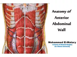
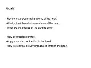
Goals: Review macro/external anatomy of the heart What is the internal/micro anatomy of the heart What are the phases of the cardiac cycle How do muscles contract Apply muscular contraction to the heart How is electrical activity propagated through the heart.

E N D
Goals: • Review macro/external anatomy of the heart • What is the internal/micro anatomy of the heart • What are the phases of the cardiac cycle • How do muscles contract • Apply muscular contraction to the heart • How is electrical activity propagated through the heart
Blood travels from the systemic system to: The right atrium ----> the right ventricle ----> pulmonary system ----> the left atrium -----> the left ventricle 6 3 1 4 5 2
Transverse section near superior border of the heart demonstrates the paricardial cavity
The heart has 3 layers • Endocardium: inner/deep layer • Myocardium: middle (muscular) layer • Epicardium: outer/superficial layer
Internal anatomy controls the direction of blood flow • Valves block “regurgitation” (Atrioventricular and Semilunar valves) • AV valves are tethered to the papillary muscles of the ventricle walls via chordae tendineae (tricuspid or bicuspid) • SL valves are more rigid, not tethered
Valves are supported by the cardiac “skeleton” Ventricles relaxed Ventricle contraction closes AV valves
The atria contract together, as do the ventricles during the cardiac cycle - Increasing heart rate shortens diastole more than systole
The atria contract together, as do the ventricles during the cardiac cycle
A heart characteristically beats in a “lubb” “dubb” pattern. The “lubb is from the AV valves closing, and the “dubb” is from the SL valves closing. During which phase of the cardiac cycle is each sound generated? Lubb - ventricle systole - isovolumetric phase/early phase Dubb - ventricle diastole - early
The microstructure of the myocardium allows for a coordinated heart beat • Cardiac muscle characteristics: • Small cells • Single nucleus • Branching, specialized connections between cells • Aerobic, have rich oxygen supply and store oxygen via myoglobin
Chapter 9! Skeletal muscle: 1 functional unit = a sarcomere - Myofilaments are surrounded by a sacroplasmic reticulum
An electric “potential” exists across muscle cell membranes • T tubules connect the sarcoplasmic reticulum to the outside of the cell • The sarcoplasmic reticulum stores Ca+ • Electric impulses (provided by nerves) travel along muscle cell membranes and induce Ca+ release into the cell
Myofilaments are overlapping in a sarcomere and slide past each other during muscle contraction
The sliding filament model of muscle contraction • Calcium is required to expose actin binding sites • ATP is required to reset the myosin head
Chapter 18 - The microstructure of the myocardium allows for a coordinated heart beat • Cardiac muscle characteristics: • Small cells • Single nucleus • Branching, specialized connections between cells • Aerobic, have rich oxygen supply and store oxygen via myoglobin
The conducting system of the heart coordinates contraction Cardiac “skeleton” between atria and ventricles prevents signal propagation from the atria to the ventricle except through the SA The sino atrial node (SA) acts as a pace maker - cells spontaneously depolarize The internodal pathways loop around both atria, trigger contraction in multiple places simultaneously The stimulus is delivered to the apex of the heart, where contractions begin in the ventricles, and are passed to other locations in the ventricles Internodal pathways converge at the atrioventricular node (AV), which conducts to the ventricles (and can also function as a pacemaker)
The relative timing of signal propagation along the conducting system and contraction in the atria and ventricles
Skeletal muscles can be over stimulated and undergo tetanus seiziure…….
….Long recovery (refractory) in the SA node pacemakers avoids this
Cardiac output (amount of blood moved by the left ventricle/minute) fluctuates to meet demand • Can increase heart rate by 250% • Can double stroke volume