Visual Pathway
Visual Pathway. Lesion 1. Lesion 2. Visual Fields and Deficits. Calcarine fissure. R. L. R. L. L. Bitemporal (heteronomous) hemianopsia. U. C. Bitemporal (heteronomous) hemianopsia. Meyers Loop (INFERIOR optic radiations, through temporal lobe ends @ inferior calcarine fissue.

Visual Pathway
E N D
Presentation Transcript
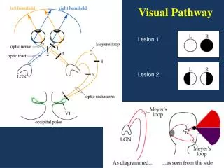
Visual Pathway Lesion 1 Lesion 2
Visual Fields and Deficits Calcarine fissure R L R L L Bitemporal (heteronomous) hemianopsia U C Bitemporal (heteronomous) hemianopsia Meyers Loop (INFERIOR optic radiations, through temporal lobe ends @ inferior calcarine fissue Left homonymous hemianopsia Left Superior quadrantanopsia Superior optic radiations, at parietal lobeends @ superior calcarine fissure Left homonymous hemianopsia w macular sparing Visual field deficits resulting from damage at different points along the primary visual pathway. The diagram on the left illustrates the basic organization of the primary visual pathway and indicates the location of various lesions. The right panels illustrate the visual field deficits associated with each lesion. (A) Loss of vision in right eye. (B) Bitemporal (heteronomous) hemianopsia. (C) Left homonymous hemianopsia. (D) Left superior quadrantanopsia. (E) Left homonymous hemianopsia with macular sparing
Optic Radiations Course of the optic radiation to the striate cortex. Axons carrying information about the superior portion of the visual field sweep around the lateral horn of the ventricle in thetemporal lobe (Meyer's loop) before reaching the occipital lobe. Those carrying information about the inferior portion of the visual field travel in the parietal lobe.