Download

1 / 19
190 likes | 301 Views
This study presents an innovative system for automated pathology classification of volumetric CT brain images, specifically focusing on traumatic brain injury (TBI) types such as extradural, subdural, and intracerebral hemorrhages. By utilizing a case-based classifier combined with sparse representation techniques, the approach eliminates segmentation and manual feature selection, significantly reducing manual workload. Experimental results demonstrate the system's effectiveness, showcasing improved utilization of medical image databases and the potential to enhance clinician decision-making through automatic image annotation.

E N D
Thien Anh Dinh1, TomiSilander1, Bolan Su1, Tianxia Gong Boon Chuan Pang2, Tchoyoson Lim2, Cheng Kiang Lee2 Chew Lim Tan1,Tze-Yun Leong1 1National University of Singapore 2National Neuroscience Institute 3Bioinformatics Institute, Singapore Unsupervised medical image classification by combining case-based classifiers
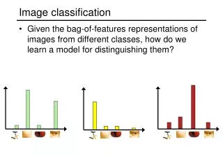
Automated medical image annotation • Huge amount of valuable data available in medical image databases • Not fully utilized for medical treatment, research and education • Medical image annotation: • To extract knowledge from images to facilitate text-based retrieval of relevant images • To provide a second source of opinions for clinicians on abnormality detection and pathology classification
Problem • Flowchart of current methods • Challenges in current methods • Highly sensitive and accurate segmentation • Extracting domain knowledge • Automatic feature selection • Time-consuming manual adjustment process • reduces usages of medical image annotation systems
Objective • An automated pathology classification system for volumetric brain image slices • Main highlights • Eliminates the need for segmentation and semantic or annotation-based feature selection • Reduces the amount of manual work for constructing an annotation system • Extracts automatically and efficiently knowledge from images • Improves the utilization of medical image databases
System overview • Case-based classifier • Gabor filters • Non domain specific features • Localized low-level features • Ensemble learning • Set of classifiers • Each classifier with a random subset of features • Final classification: an aggregated result
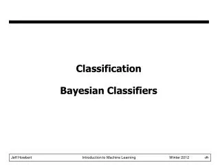
Sparse representation-based classifier • Sparse representation-based classifier (SRC) proposed by Wright et al. for face recognition task • Non-parametric sparse representation classifier • SRC consists of two stages • Reconstructing: a test image as a linear combination of a small number of training images • Classifying: evaluating how the images belonging to different classes contribute to the reconstruction of the test image
Image databases x1, x2,…, x1000 Sparse reconstruction New data item y ≈ a7x7 + a23x23 + a172x172 + a134x134 + a903x903 y ≈ a7x7 + a23x23 + a172x172 + a134x134 + a903x903 Class residuals r1 = || y – (a7x7 + a172x172 + a132x134)||2 r2 = || y – (a23x23 + a903x903)||2
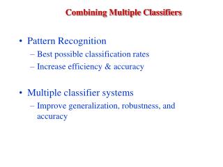
Ensemble of weak classifiers • Combine multiple weak classifiers • Take class specific residuals as confidence measures • The smaller the residual for the class, the better we construct the test by just using the samples from that class • To classify image y, compute average class-specific residuals of all W weak classifiers
Domain • Automatically annotate CT brain images for traumatic brain injury (TBI) • TBI: major cause of death and disability • Several types of hemorrhages: • Extradural hematoma (EDH) • Subdural hematoma (SDH) • Intracerebralhemorrage (ICH) • Subarachnoid hemorrhage (SAH) • Intraventricular hematoma (IVH) Extradural hematoma Subdural hematoma
Data • CT brain scans of 103 patients • Each scan: • Volumetric stack of 18-30 images (slices) • Image resolution: 512 x 512 pixels • Manually assigned a hematoma type extracted from its medical text report
Experimental setup • Compared performances of • SRC vs. SVM vs. SVM + feature selection • With/without ensemble learning • Run stratified ten-fold cross-validation 50 times with different random foldings • Measured the average precisions and recalls • Separated training and testing dataset at the case level
Experimental results when varying the ensemble size Average precision and recall of classifiers when varying the ensemble size (number of features = 1000)
Experimental results when varying the number of features per classifier Average precisions and recalls of classifiers when varying number of features (ensemble size = 50)
Conclusion • Ensemble classification framework with sparse Gabor-feature based classifier • Eliminates the requirement for segmentation and supervised feature selection • Reduces the need for manual adjustment • Achieves reasonable results compared to segmentation dependent techniques (Gong et al.) • Limitation • Longer classification time when dealing with large training data • Manual weighting needed for imbalanced data
Gabor features • Localize low level features from an input image • Resemble the primitive features extracted by human visual cortex • Extract edge like features in different scales and orientations at different locations of the image • Create a Gabor filter bank with 5 frequencies and 8 orientations • A 128 x 128 grayscaleimage: 655360 features • Randomly select 4000 Gabor features to form a feature subspace