Activity-Based Protein Profiling
Activity-Based Protein Profiling. Contents. Introduction -Assignment of Protein Function In the Postgenomic Era -Detection Strategies for Activity-Based Proteomics -What Is an Activity-Based Probe Application of Activity-Based Probes -Identification of Biomarkers for Human Disease
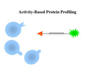

Activity-Based Protein Profiling
E N D
Presentation Transcript
Contents Introduction -Assignment of Protein Function In the Postgenomic Era -Detection Strategies for Activity-Based Proteomics -What Is an Activity-Based Probe Application of Activity-Based Probes -Identification of Biomarkers for Human Disease -In Vivo Imaging of Enzyme Activities -Small Molecule Screening and Target Discovery Case-discussion -p90 ribosomal protein S6 kinases -Metalloprotease -Cysteine protease Conclusion
Assignment of protein function in the postgenomic era * In the postgenomic era researchers are now confronted with the task of assigning functions to tens of thousands of proteins. Many proteins, such as enzymes, are functionally regulated by a series of post- translational mechanisms, leading to a lack of correlation between activity and expression levels. Global analysis of changes in gene transcription and translation by abundance-based genomic and proteomic approaches provides only indirect information about protein function. Activity-based protein profiling (ABPP) is a chemical strategy that utilizes active site directed covalent probes to profile the functional state of enzymes in complex proteomes.
Detection strategies for activity-based proteomics * Unraveling the functional roles of proteins is a major challenge facing the post-genome researcher Advances towards this goal have been made through the development of both chemical and biochemical tools for monitoring protein activity Examples (3 becomes more popular now) 1. Small-molecule substrate reporters of enzymatic activity 2. Protein-based reporters of enzymatic activity 3. Activity-based probe
Small-molecule substrate reporters of enzymatic activity * These reagents carry fluorescent groups, and thus energy emission upon their enzymatic conversion to product can be monitored over time Disadvantages: 1.The majority of basic fluorogenic probes cannot be directly applied to complex cellular environments 2. The another challenge in using the approach lies in the ability to generate probes that are specific for an individual enzyme (a peptide has the potential to function as a substrate for more than one class of proteolytic enzymes) TRENDS in Cell Biology, Vol.14 No.1 January 2004
Protein-based reporters of enzymatic activity Fluorescent reporters Bioluminescent reporters Disadvantages: 1.The use of FRET has been extended further to design biochemical tools for monitoring enzymatic activity inside cells 2. They all suffer from the selectivity of probes for a specific enzyme target TRENDS in Cell Biology, Vol.14 No.1 January 2004
What Is an Activity-Based probe (ABP) ? * The activity-based probes (ABPs): they generally contain three main functional groups: 1. The chemical reactive group or warhead (covalently modifies an active-site residue of the enzyme of interest) 2. A linker region, which can be specific for different enzymes 3. A tag, which is used to visualize the modified enzyme Warhead linker tag
The reactive group of activity-based probe * The reactive group is perhaps the most significant and difficult piece of the probe to design. It functions to covalently link the ABP to an amino acid residue in the target enzyme’s active site when the target enzyme is active. Nature chemical biology, Vol.1 No.3 August 2005
The reactive group of activity-based probe (2) Nature chemical biology, Vol.1 No.3 August 2005
The mechanism of reactive group for enzyme targets Chemical Reviews, Vol. 106, No.8, 2006
The tag region of activity-based probe * The tag allows the identification or purification of modified enzymes. Biotin, fluorescent small molecules, and radioactive isotopes are most commonly incorporated into ABPs as tags Current Opinion in Chemical Biology, Vol.11, 2007
The linker region of activity-based probe * The linker region can be viewed as a bridge between the reactive group and the labeling tag. The linker serves to prevent steric hindrance by the tag that could inhibit the reactivity of the probe, a linker can take the form of an extended alkyl or polyethylene glycol (PEG) spacer. • The linker can serve as a specificity factor enabling targeting of the probe to a specific enzyme or class of enzymes. For example , to target proteases, this specificity region can be engineered to contain peptide sequences.
Application of Activity-Based Probes with affinity tag * Identification of Biomarkers for Human Disease Am. J. Pharmacogenomics , Vol.4, No.6 2004
Application of Activity-Based Probes with fluorescent tags * In Vivo Imaging of Enzyme Activities Am. J. Pharmacogenomics , Vol.4, No.6 2004
Competition of Activity-Based Probes with inhibitors * Small Molecule Screening and Target Discovery Am. J. Pharmacogenomics , Vol.4, No.6 2004
Case-discussion * p90 ribosomal protein S6 kinases
RSK and MSK in MAP kinase signalling RSK (Ribosomal protein S6 Kinase) and MSK (Mitogen- and Stress- activated protein Kinase) constitute a family of protein kinases that mediate signal transduction downstream of MAP kinase cascades. RSK is activated by MAP kinases of the extracellular signalregulated kinase (ERK) family in response to growth factors, many polypeptide hormones, neurotransmitters, chemokines and other stimuli.
The domain structure and activation of RSK Domain structure Activation and inactivation • The N-terminal kinase domain (NTK) belongs to the AGC kinase family and is responsible for phosphorylation of substrates. • The C-terminal kinase domain (CTK) belongs to the CamK family and its only known function is activation of NTK. *AGC:containing PKA, PKG, PKC kinases family *CamK: calmodulin-dependent protein kinase family J. Cell Sci.119, 3021–3023 (2006)
Structural bioinformatics-based design of selective, irreversible RSK inhibitors Previous targeting strategy on ATP-binding site • All kinase inhibitors target the adenosine triphosphate (ATP) binding site • The ATP binding sites of 491 human protein kinase domains are highly conserved, which makes the design of selective inhibitors a formidable challenge. • Structural bioinformatics approach to identify two selectivity filters: a threonine and a cysteine, at defined positions in the active site of p90 ribosoma lprotein S6 kinase (RSK) Unique design targeting non-conserved regions Science308, 1318–1321 (2005)
RSK inhibitor X Structural bioinformatics-based design of selective, irreversible RSK inhibitors • Selectivity filter 1: compact gatekeeper---Threonine • allows bulky aromatic substituents, such as those found in the Src family kinase inhibitors, PP1 and PP2, to enter a deep hydrophobic pocket as ~20% of human kinases have a threonine at this position • Selectivity filter 2: chemical reactive amino acid--- Cysteine • Out of 491 related kinase domains in the human genome, there are 11 kinase with a cysteine at the C-terminal end of the glycine-rich loop • A cysteine near this solvent exposed loop is likely to have a lower pKa and therefore to be more reactive than a cysteine buried in the hydrophobic pocket Science308, 1318–1321 (2005)
p-tolyl substituent Reactive group: Fluoromethylketone (fmk) Chloromethylketone (cmk) RSK RSK Cell-based assay (for HEK-293 cell) In vitro assay (for RSK2) PMA: Phorbol Myristate Acetate * IC50 in uM * EC50 of ~150 nM Structural bioinformatics-based design of selective, irreversible RSK inhibitors Hydrophobic packet Reactive cysteine Science308, 1318–1321 (2005)
Fluorescent tag The design of an fmk derivative (1) * EC50 of >10 uM Oncogene25, 5764–5776 (2006)
* Click chemistry method Chemistry & Biology. 11, 535-546 (2004) The design of an fmk derivative (2)
A clickable inhibitor for RSK TAMRA is a fluorescent azide * EC50 of ~30 nM
Discussion • fmk-pa, a propargylamine variant that has improved cellular potency and a ‘clickable’ tag for assessing the extent and selectivity of covalent RSK modification. • Saturating concentrations of fmk-pa inhibited Ser386 phosphorylation and downstream signaling in response to phorbol ester stimulation, but had no effect on RSK activation by lipopolysaccharide. • Clickable inhibitors such as fmk-pa should facilitate determination of the specific roles played by the RSK CTD in cellular and animal models relevant to heart failure and other human diseases.
Case-discussion (2) * Metalloprotease Metalloproteases are a large, diverse class of enzymes involved in many physiological and disease processes.
Metalloprotease (requiring activator) • Metalloproteases are regulated by post-translational mechanisms that diminish the effectiveness of conventional genomic and proteomic methods for their functional characterization . inactive precursor enzyme (zymogens) endogenous binding proteins (TIMPs)
AOMK • For cysteine protease : Acyloxy methyl ketone (AOMK) group
L L Metalloprotease Hydroxamate (Hx) group Hx: zinc-chelating group (non-covalent bond) Activity-based Probes Design of Metalloprotease • For metalloprotease : • do not use a catalytic amino acid side chain as the primary nucleophile • catalytic zinc ion PNAS 101, 10000-10005 (2004)
Rhodamine Hx group benzophenone (BP): photo-cross-linker (for covalent bond formation) Activity-based Probes Design of Metalloprotease • First generation metalloproteases ABPs:
Click chemistry method Chemistry & Biology. 11, 535-546 (2004) Activity-based Probes Design of Metalloprotease • New generation metalloproteases ABPs: The large reporter tag which might be expected to obstruct interactions with certain metalloproteases
* General structure of the alkyne-tagged hydroxamate-benzophenone (HxBPyne) probe Activity-based Probes Synthesis of Metalloprotease
Mouse liver proteome Proteomic profiling of the HxBPyne probe library
Proteomic profiling of the HxBPyne probe library * Recombination expression sample (breast cancer)
4 µg/ml of MMP in a background of 1 mg total protein / ml 1 µM probe Proteomic profiling of the HxBPyne probe library * Detection limit 1 uM LeuR2 HxBPyne
MudPIT Profiling Metalloproteases Activities by ABPP-MudPIT • ABPP-MudPIT : Activity-Based Protein Profiling with Multidimensional Protein Identification Technology For enhancement of resolution and sensitivity MudPIT Nat. Bioltechnol. 19, 242-247 (2001)
Sensitivity of Detection of MMPs by ABPP-MudPIT 100 nM of the LeuR2 HxBPyne probe and analyzed by ABPP-MudPIT C: LeuR2 HxBPane (control) detection limit 0.001~0.01% ( 5~50 fold)
Profiling Metalloprotease Activities in Cancer proteome • To identify endogenous metalloprotease activities and quantify their relative levels in disease states the optimal probe set (cocktail): 100 nM of each HxBPyne probe; total 400 nM total probe Invasive Non-Invasive melanoma *C: 100 HxBPane competitor probes
Discussion (2) • ABPP may facilitate the simultaneous discovery of enzyme activities associated with human disease and chemical tools for testing their function in pathological processes
Quencher Case-discussion (3) * Cysteine protease: cathepsins
Cathepsins are usually characterised as members of the lysosomal cysteine protease (active site) family. • Elevated cathepsin enzyme activity in serum or the extracellular matrix often signifies a number of gross pathological conditions. • Cathepsin-mediated diseases include: Alzheimer's, numerous types of cancer, autoimmune related diseases like arthritis and the accelerated breakdown of bone structure seen with osteoporosis Papain-family protease (cathepsin B and L) Chemical Reviews, 2002, Vol. 102, No. 12
Cysteine cathepsins in human cancer Biol. Chem., Vol. 385, pp. 1017–1027, November 2004
2,6-dimethyl benzoic acid AOMK N-protected glycine AOMK AOMK: Acyloxy methyl ketone (warhead) F K BODIPY ( tag) AOMK Synthesis of the qABP GB117 and the control ABP GB111 Phenylalanine-lysine dipeptide (linker) ABP
BODIPY ( tag) G QSY7 (quencher) QSY7 (quencher) Synthesis of the qABP GB117 and the control ABP GB111 qABP
Determination of quenching efficiency of qABP GB117 relative to the unquenched control GB111 LysoTracker (lysosomal marker): Weakly basic amines selectively accumulate in cellular compartments with low internal pH and can be used to investigate the biosynthesis and pathogenesis of lysosomes
Structure of the new qABPs (NIRF-ABPs) The most stable probe
* Inhibitor: GB111-NH2 Labeling of recombinant cathepsins and intact cells with the control ABP and qABP Non- specific * NIH-3T3 cells