Download
1 / 40
410 likes | 605 Views
Growth and Repair. dr. FAIRUZ QUZWAIN,SpPA,M.Kes Kepala bagian Patologi Anatomi FKIK-UNJA. Repair following inflammation: two simultaneous processes. Regeneration : replacement of injured/necrotic cells by cells of same type, often leaving no evidence of previous injury

E N D
Growth and Repair dr. FAIRUZ QUZWAIN,SpPA,M.Kes Kepala bagian Patologi Anatomi FKIK-UNJA
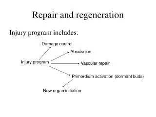
Repair following inflammation: two simultaneous processes • Regeneration : replacement of injured/necrotic cells by cells of same type, often leaving no evidence of previous injury • Repair : replacement of injured/necrotic cells by connective tissue, leaving a permanent scar (microscopic or macroscopic)
Cell Cycle PHASES of Cell Cycle: G1: S: G2: M: G0:
Correlation of Cell Cycle and Tissue Types • Continuously dividing (labile) cells: • Surface epithelium and excretory ducts of glands (skin, gi / gu mucosa, biliary tract, pancreas) • Marrow hematopoietic cells • Stem cells in multiple organs (immature, undifferentiated cells) • Quiescent (stable) cells in G0: • Organ parenchymal cells (liver, kidneys) • Mesenchymal cells (fibroblasts, smooth muscle, endothelium, chondrocytes, osteocytes) • Nondividing permanent cells (can’t re-enter cell cycle) • Neurons, skeletal & cardiac myocytes
Mechanism: control of cell cycle Which cyclin ? Which cyclin? Cyclin-dependent kinase inhibitors
Modes of Intercellular Signalling Autocrine Paracrine Endocrine
Surface Receptors: 3 classes Receptors without intrinsic tyrosine kinase Receptors with intrinsic tyrosine kinase Seven transmembrane (G protein-coupled)
Consequences of Receptor Activation • Intrinsic-kinase activity receptors: • Irreversible commitment of cell to enter (proliferative response) • Receptors without intrinsic kinase activity (cytokine superfamily): • Activation cytosolic kinases to mediate functional response (not proliferative) • G-protein coupled (seven spanning) receptors: • Over 1500 receptors identified • Bind various ligands, producing specific intracellular response
Signal Transduction by Tyrosine Kinase Receptors Growth factors: coming soon! Clinical application: if mutant ras protein is permanently fixed in active GTP form, what pathologic process may result?
Transcription Factors • Definition: intracellular proteins that regulate gene expression, thereby controlling cell growth • Specific domains in transcription factors: • : permits factor to bind specifically to short sequences of DNA • : allows factor to increase transcription of DNA • :allows factor to decrease transcription of DNA • Transcription factors known to be operative in malignant neoplasms: • Growth promoting: c-MYC and c-JUN • Cell cycle inhibiting (tumor suppressor gene): p53
Growth Factors • Definition: proteins that bind to cell surface receptors with generating cascade response that signals cell to enter S-phase (cell division). • These factors can also modulate cell functions: locomotion, contractility, differentiation, etc.
Major growth factors / effects EFFECTS • Mitogenic for epithelium & fibroblasts • Mitogenic for hepatocytes Mitogenic for endothelial cells • Mitogenic for monocytes, fibroblasts, smooth muscle cells; activates neutrophils • Angiogenesis, wound repair (mitogenic for both fibroblasts and keratinocytes) FACTOR • EGF = epidermal • TGF-a = transforming • VEGF=vascular endothelial • PDGF= platelet-derived • FGF= fibroblast • FGF-1=acidic • FGF-2=basic
Tissue Regeneration Liver from living donor before transplantation, outlining right lobe to be used for grafting into recipient Liver of donor one week post-partial hepatectomy, showing marked growth of left lobe (compensatory hyperplasia) without regrowth of right lobe. Why didn’t right lobe regrow also?
Extracellular matrix 1 • Definition: macromolecules outside cells, formed by local secretion and assembled into network surrounding cells • Functions: • Sequester H2O for turgor; minerals for rigidity • Reservoir for growth factors • Scaffolding within which cells adhere, migrate, and proliferate
Extracellular matrix (ECM) 2 • Groups of macromolecules in ECM: • Fibrous structural proteins: 2 major families are: • Adhesive glycoproteins • Gel proteins in intercellular junctions and cell surfaces: proteoglycans & hyaluronic acid
Extracellular matrix (ECM) 3 • Macromolecules of ECM assemble into two types of organizational structure: • : fills spaces between cells • : closely associated with cell surfaces
Collagen: summary of major types Skin (80%), bone (90%), tendons Genetic deficiency of type IV in:
Collagen synthesis Nutrient required for hydroxylation of alpha chains: Deficiency of this nutrient causes poor wound healing in disease called: Inherited disorders of collagen synthesis, leading to defective fibers:
Elastic Fibers • Definition: fibers capable of stretching and recoiling to original size • Present in tissues requiring elasticity: • Structure: • Central core protein: • Peripheral microfibrillary network: • Inherited defect synthesis of peripheral microfibrillary network: abnormally weakened elastic fibers. Syndrome? Skin, lung, uterus, ligaments, large blood vessels
Adhesion molecules 1 Function: attach cells to ECM matrices; 2 glycoprotein chains held together by disulfide bonds; produced by fibroblasts, endothelial cells, & monocytes. Name?
Adhesion molecules 2 Most abundant glycoprotein in basement membranes; it spans basal lamina and binds to both cell surfaces and ECM components:
Adhesion molecules 3 Transmembrane glycoproteins with alpha and beta chains that bind to fibronectin, laminin, & collagen. This family of surface receptors mediate attachment of cell membranes to ECM: These also mediate adhesion of which cell type to endothelium?
Summary: interactions cell-ECM Major EC structural protein: Fig. 3-16, Pathologic Basis of Disease, 7th ed, Elsevier 2005
Overview: Repair after injury ACUTE AND CHRONIC INFLAMMATION Damage to parenchymal cells and interstitial framework Regeneration of parenchymal cells whenever possible Replacement of non-regenerated damaged tissue by what?
Fibrosis (fibroplasia) • Four components: • : formation new blood vessels • of fibroblasts into damaged tissue • of extracellular matrix • Organization fibrous tissue =
Sequence of events in repair 24 hrs: proliferation of fibroblasts & endothelial cells Within 3-5 days: Permanent result (weeks later) Little mature collagen Proliferation of young fibroblasts blue-staining collagen (trichrome stain) New capillaries
Angiogenesis • Definition: pre-existing vessels send out capillary sprouts to form new vessels • cf. vasculogenesis: the primitive vascular network established during embryogenesis • Clinical importance: • Repair post-inflammation • Formation collateral circulation (post-MI) • Support growth of neoplasms (therapeutic implications)
ECM proteins affecting angiogenesis • Integrins: formation and maintenance new vv. • Matrix proteins which destabilize cell-matrix interactions, promoting angiogenesis: • Thrombospondin • SPARC • Tenascin C • Proteases that remodel matrix • Plasminogen activators • Matrix metalloproteinases • Fragment of collagen that inhibits endothelial proliferation and angiogenesis, with therapeutic application in neoplasia?
Fibrosis (fibroplasia) • Emigration and proliferation of fibroblasts at injury site, triggered by multiple growth factors produced by cells in granulation tissue, most important of which is: • ECM deposition by fibroblasts: fibrillar collagen synthesis enhanced by growth factors and cytokines, thus converting Into a
Tissue remodeling • Conversion granulation tissue into scar involves changes in composition of ECM. • : enzymes which degrade ECM components for remodeling. These enzymes are dependent on ions for activity.
Wound Healing Healing by first intention Healing by second intention
Summary: phases of wound healing Wound tensile strength: 10% of normal at 7 days; 70-80% of normal at 3 months
Factors influencing wound healing • Local Factors • : most important single cause of delay • Mechanical: too early motion can delay • Foreign bodies: may impede or cause abscess • Location: speed of healing proportional to richness of blood supply: face > trunk > extremities • Type of wound: primary intention heals faster than secondary intention
Pathologic complications, 2 • Excessive formation of repair components: • Excessive granulation tissue • Desmoid tumor (aggressive fibromatosis) • Best viewed as low grade neoplasm with stubborn tendency for recurrences
Conclusion • Physicians stand in wonder at the amazing capacity of the body to restore itself after injury, usually without loss of normal function. • This represents an advanced kind of engineering and self-regulated maintenance function that humbles human technology.