Download
1 / 34
370 likes | 613 Views
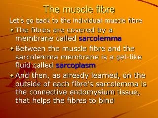
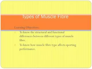
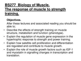
BS277 Biology of Muscle. Fibre Types. Objectives. After this lecture and associated reading you should be able to; Discuss the relative contributions of genetics and environment to muscle fibre isotype. Discuss the molecular basis of fibre isotype structure and function.

E N D
BS277 Biology of Muscle.Fibre Types Objectives. After this lecture and associated reading you should be able to; • Discuss the relative contributions of genetics and environment to muscle fibre isotype. • Discuss the molecular basis of fibre isotype structure and function.
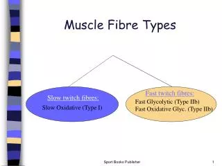
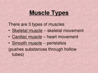
Twitch characteristics of different (phasic) muscles reflect fibre type composition. Note that most muscles contain mixtures of fast (majority) and slow fibres.
As a consequence of twitch time course, a fast muscle requires a higher frequency of stimulation to develop tetanus tension. Force generated during 500-ms stimulation at different frequencies (isolated mouse muscles, 25°C). A, soleus; B, extensor digitorum longus (EDL).
TABLE 10-3Properties of Skeletal Muscle Fibre Types Property Slow Intermediate Fast Contraction speed (Tension) Fatigue resistance Colour Myoglobin content Capillary supply Mitochondria [Glycolytic enzyme] Substrates used for ATP generation
TABLE 10-3Properties of Skeletal Muscle Fibre Types Property Slow Intermediate Fast Contraction speed Slow Fast Fast (Tension) Fatigue resistance Colour Myoglobin content Capillary supply Mitochondria [Glycolytic enzyme] Substrates used for ATP generation
TABLE 10-3Properties of Skeletal Muscle Fibre Types Property Slow Intermediate Fast Contraction speed Slow Fast Fast (Tension Low Intermediate High) Fatigue resistance Colour Myoglobin content Capillary supply Mitochondria [Glycolytic enzyme] Substrates used for ATP generation
TABLE 10-3Properties of Skeletal Muscle Fibre Types Property Slow Intermediate Fast Contraction speed Slow Fast Fast (Tension Low Intermediate High) Fatigue resistance High Intermediate Low Colour Myoglobin content Capillary supply Mitochondria [Glycolytic enzyme] Substrates used for ATP generation
TABLE 10-3Properties of Skeletal Muscle Fibre Types Property Slow Intermediate Fast Contraction speed Slow Fast Fast (Tension Low Intermediate High) Fatigue resistance High Intermediate Low Colour Red White White Myoglobin content Capillary supply Mitochondria [Glycolytic enzyme] Substrates used for ATP generation
TABLE 10-3Properties of Skeletal Muscle Fibre Types Property Slow Intermediate Fast Contraction speed Slow Fast Fast (Tension Low Intermediate High) Fatigue resistance High Intermediate Low Colour Red White White Myoglobin content High Low Low Capillary supply Mitochondria [Glycolytic enzyme] Substrates used for ATP generation
TABLE 10-3Properties of Skeletal Muscle Fibre Types Property Slow Intermediate Fast Contraction speed Slow Fast Fast (Tension Low Intermediate High) Fatigue resistance High Intermediate Low Colour Red White White Myoglobin content High Low Low Capillary supply Dense Intermediate Scarce Mitochondria [Glycolytic enzyme] Substrates used for ATP generation
TABLE 10-3Properties of Skeletal Muscle Fibre Types Property Slow Intermediate Fast Contraction speed Slow Fast Fast (Tension Low Intermediate High) Fatigue resistance High Intermediate Low Colour Red White White Myoglobin content High Low Low Capillary supply Dense Intermediate Scarce Mitochondria Many Intermediate Few [Glycolytic enzyme] Substrates used for ATP generation
TABLE 10-3Properties of Skeletal Muscle Fibre Types Property Slow Intermediate Fast Contraction speed Slow Fast Fast (Tension Low Intermediate High) Fatigue resistance High Intermediate Low Colour Red White White Myoglobin content High Low Low Capillary supply Dense Intermediate Scarce Mitochondria Many Intermediate Few [Glycolytic enzyme] Low High High Substrates used for ATP generation
TABLE 10-3Properties of Skeletal Muscle Fibre Types Property Slow Intermediate Fast Contraction speed Slow Fast Fast (Tension Low Intermediate High) Fatigue resistance High Intermediate Low Colour Red White White Myoglobin content High Low Low Capillary supply Dense Intermediate Scarce Mitochondria Many Intermediate Few [Glycolytic enzyme] Low High High Substrates used for Lipids, CHO, Primarily CHO ATP generation aas (aerobic) CHO (anaerobic) (anaerobic)
TABLE 10-3Properties of Skeletal Muscle Fibre Types Property Slow Intermediate Fast Contraction speed Slow Fast Fast (Tension Low Intermediate High) Fatigue resistance High Intermediate Low Colour Red White White Myoglobin content High Low Low Capillary supply Dense Intermediate Scarce Mitochondria Many Intermediate Few [Glycolytic enzyme] Low High High Substrates used for Lipids, CHO, Primarily CHO ATP generation aas (aerobic) CHO (anaerobic) (anaerobic) Alternative names Type I, S (slow), Type IIa, FOG Type IIb/x, SO (slow oxidative) FR (fast resistant) FAG, FF (Fast fatigue)
The molecular basis for fibre isotype differences. 2xMHC 200kD 4xMLC 20kD S1 ATPase and actin binding. S2 aggregation and hinge. 300 mols per thick filament (Regulatory) MHC (essential) Thin myofilament is actin + regulatory proteins
The myosins from different species with different speeds of muscle contraction break down ATP at different rates. i.e. the contraction speed of muscle depends on the enzyme activity of the actomyosin. Fast muscle uses ATP quickly. From Barany, 1967. In Jones et al 2004
The ATPase activity of the myofibril is a property of the kind of myosin it contains. Isotypic variation in myosin • Myosins within species also differ between fibres with different shortening velocities. • Human has 4 heavy chain isotypes. • Type 1 (slow) • Type 2 (fast) • Type 2a • Type 2x (b) • Type 2c (Embryo and regenerating fibres)
Histochemical identification of myosin isotypes. • Type 1 (slow) inactivated by preincubation at pH 9.4 • Type 2 (fast) • Type 2a inactivated by pre-incubation at 4.3-4.6 • Type 2x (b) activity reduced by pre-incubation at 4.3-4.6 • Type 2c (Embryo and regenerating fibres)
Isotypic variation in myosin expression • Imunohistochemistry can detect structural differences between MHC isotypes. • About 3% of fibres express both type 1 and type 2 MHC. • Up to 40% can express both 2a and 2x. • 2 light chains for each heavy chain; essential and regulatory (only regulate in smooth muscle). • Light chains also have fast and slow isotypes.
Isotypic variation in other myofibrillar proteins • Actin has skeletal and cardiac forms but no differential association with fast / slow fibre isotypes. • Tropomyosin is an αβ dimer. • Can be homo or hetero dimeric (αα; ββ; αβ) • α chain has fast and slow forms. • Fast fibres tend to have ββ and the fast form of α • Troponin • TnC, TnI(inhibitory), TnT(tropomyosin binding) • All have fast and slow forms. Schaffiano, S. and Reggiani, C. (1996) Physiol. Rev 76, 371-421.
Methods of fibre typing • Biopsies in humans; cryostat sections cut. • Enzyme histochemistry • myosin ATPase • Fast; inactivated at pH 4.3-4.6. • Slow; inactivated by pH 9.4. • ‘glycolytic’ enzymes • LDH • -glycerophosphate dehydrogenase • PFK • glycogen (myo)phosphorylase • ‘aerobic’ enzymes • Kreb’s cycle • Succinate dehydrogenase (SDH) • Lipid / ketone metabolising enzymes • beta hydroxybutyrate dehydrogenase • Non-specific esterase • Immunohistochemistry • Myosin heavy/light chains
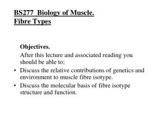
Identification of human skeletal muscle fibre isotype by enzyme histochemistry. (From Jones et al 2004) Micrograph shows sequential transverse sections of quadriceps stained for myosin ATPase (pH 9.4; inactivates type 1 myosin)), NADH transferase (complex 1; oxidative) and (myo)phosphorylase.
Identification of human skeletal muscle fibre isotype by enzyme histochemistry.(From Jones et al 2004) Enzyme profiles from serial sections stained using different substrates, identify clusters of properties that differentiate between fibre types. SDH = succinate dehydrogenase (‘aerobic’) -GPDH = glycerophosphate dehydrogenase (glycolytic)
Proportions of Type 1 (slow) fibres in different populations (From Spurway, 2006)
Contractile characteristics of the main types of motor unit. Upper traces are single twitches; lower traces show the fatigue curves during repetitive tetanic stimulation. Note the differences in force, speed and fatigability. Tension (One stimulus) (Repeated stimuli)
( number of fibres) Contractile properties of motor units. The size, speed and fatigue resistance of individual motor units, correlates with fibre type as defined by histochemical properties.
Neural determination of fibre isotype. • Similarity of fibres within a unit suggest a neural influence. • Cross-innervation changes phenotype (Buller et al, 1960). • Denervated/tenotomised postural muscle becomes faster • Electrical stimulation at 10Hz type 1. • Brief, infrequent 40 Hz type 2X • More frequent 40 Hz type 2A • Frequent stimulation elevates Ca++ that regulates transcription factor (NFAT) activity via calcineurin and NFAT dephosphorylation. (Chin, 1998) • Inheritance of fibre type via motor neurones?
Other factors. • If the nervous system does not develop, fibre types still differentiate. • Steroids effect fibre size, but [thyroid hormone] influences fibre type (higher T3/4, faster isoforms). • Interaction between muscle activity and hormones. More active muscles more susceptible to hormone influence • Stretch ( tension) fast-slow transformation.
Overview of differences between athletes. • % of type 1:type 2 determined genetically (motor neurones? Myoblasts?). • Proportions of type 2A and type 2X influenced by training • (high frequency of recruitment induces 2X 2A, but not type 2 type 1). • Frequency of recruitment influences hypertrophy (type 2 > type 1). • Frequency of recruitment influences levels of metabolic enzymes. • Training intensity and volume influence hormones; synergistic effect with direct influence on muscles?
Luquet, S. et al.(2003). Peroxisome proliferator-activated receptor δ controls muscle development and oxidative capability. FASEB J. 17, 2299-2301 PPAR is a ligand activated transcription factor.