Download
1 / 33
330 likes | 410 Views
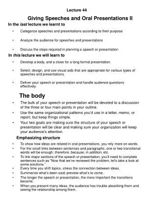
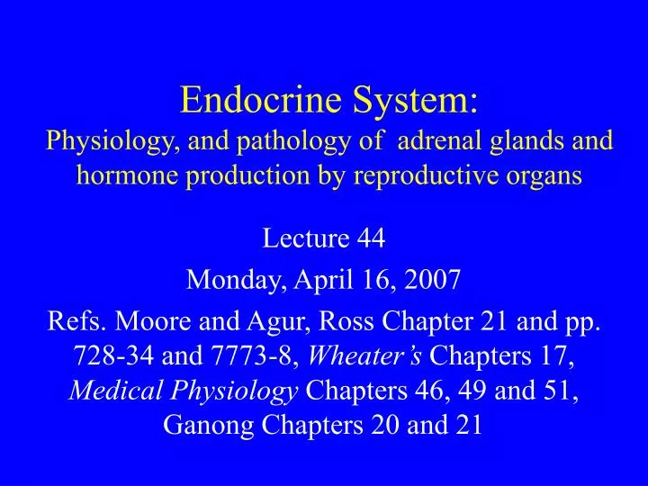
Endocrine System: Physiology, and pathology of adrenal glands and hormone production by reproductive organs. Lecture 44 Monday, April 16, 2007

E N D
Endocrine System:Physiology, and pathology of adrenal glands and hormone production by reproductive organs Lecture 44 Monday, April 16, 2007 Refs. Moore and Agur, Ross Chapter 21 and pp. 728-34 and 7773-8, Wheater’s Chapters 17, Medical Physiology Chapters 46, 49 and 51, Ganong Chapters 20 and 21
Control of secretion of cortisol. Solid arrows are stimulatory effects. Dashed arrows show inhibition. Ganong 20-21
Prolonged high glucocorticoid levels • Chronic stress (emotional or physical) can override negative feed back of serum cortisol and result in prolonged high endogenous glucocorticoid levels. • Deleterious effects include: • Tissue wasting, hyperglycemia, hypertension, delayed wound healing. • Atrophy of lymphoid tissue. Lymphopenia. Decreased immune function. Increased risk of infection.
Adrenocortical hyperfunction • 1. Cushing’s syndrome • Excess glucocorticoids, endogenous cortisol or potent corticosteroid drug • 2. Hyperaldosteronism • Excess aldosterone • 3. Adrenogenital syndromes • Usually excess androgens
Causes of Cushing’s syndrome • 1. Exogenous steroids--Iatrogenic. • Because corticosteroids are used to treat many inflammatory conditions, this is the most common form clinically. • Corticosteroid administration has negative feedback on hypothalamus and pituitary. • Lack of ACTH secretion results in atrophy of adrenal cortex. • Treatment involves withdrawing drug slowly. • Abrupt termination can cause adrenal crisis. • Endogenous forms(3 types)
Causes of Cushing’s syndrome • 2. Hypersecretion of ACTH >50% of endogenous cases (Known as Cushing’s Disease) • Usually due to pituitary adenoma. • High serum ACTH and bilateral cortical hyperplasia. • 3. Primary adrenal secretion 25-30% of endogenous cases • Usually due to a functional adenoma or adeno-carcinoma of the adrenal cortex. • Low serum ACTH and often non-neoplastic cortex is atrophied • 4. Paraneoplastic syndrome • Secretion of ACTH by neoplasm of tissue that does not normally produce ACTH, e.g. small cell carcinoma of lung.
Laboratory results in endogenous Cushing syndrome BP Table 20-7
Hormone synthesis in the zona glomerulosa. This zone lacks 17a-hydroxylase and is the only zone that has aldosterone synthase.A II is angiotensin II.Ganong 20-9
Effects of mineralocorticoids • Increase the reabsorption of Na+ from: • urine, sweat, saliva colonic contents • Act mainly on P cells of collecting ducts • exchange Na+ for K+ and H+ • Mechanism: binds cytoplasmic receptor and moves to nucleus and alters transcription • increase ENaC insertion into membrane • increase ENaC synthesis • possible direct effect in increasing Na+-K+ exchangers
Hyperaldosteronism • Excessive secretion of aldosterone causes Na+ retention and K+ loss resulting in hypertension and hypokalemia. • Primary- Adrenal hyperfunction • Serum renin and angiotensin II will be low. • Usually due to primary adenoma- Conn’s syndrome. • May be caused by primary adrenocortical hyperplasia • Secondary- due to activation of renin-angiotensin system. • Angiotensin II stimulates aldosterone secretion. • Examples: Congestive heart failure, renal nephrosclerosis, hypoalbuminemia
Adrenogenital syndrome • Excess androgen secretion causes virilizing effects • Primary gonadal disorder • Adrenal secretion of excess androgens: • Neoplasm of adrenal cortex. • Deficiency of enzyme necessary for cortisol synthesis (Low cortisol induces ACTH and ACTH causes hyperplasia and increased androgen secretion )
Normal (top) and nodular hyperplasia of adrenal cortex. PBD 6th ed 26-26
Hypoadrenocorticism • Mineralocorticoids and glucocorticoids are essential for life • Addison’s disease is primary adrenal insufficiency due to destruction of the adrenal cortex • Now usually caused by autoimmune destruction • Formally often caused by tuberculosis • Clinical signs: hypotension, fatigue, weight loss • Clinical pathology: low Na+, high K+ • Fatal due to shock, if untreated. • Secondary adrenocortical insufficiency • Due to lack of ACTH • Deficient cortisol and androgens
Adrenal medulla • In human 90% of medullary cells secrete epinephrine and 10% secrete norepinephrine. • Adrenal medullary hormone secretion is not essential for life. • Adrenal medullary hormones help deal with emergencies. • Most of the catecholamines in plasma come from the medulla. • Metabolites of secreted catecholamines are excreted in the urine.
Diseases of the adrenal medulla • The most important diseases are neoplasms: • Pheochromocytoma- uncommon • Tumor of chromaffin cells. • Can cause hypertension (surgically correctable) • Hypertension can be fatal when a stressful stimulus causes massive secretion of catecholamines. • Benign or malignant • Neuroblastoma • Originate anywhere in sympathetic nervous system • Most common extracranial tumor in children
Pheochromocytomatumor of adrenal medulla PBD 6th ed. 26-33see also BP 20-27
Hormone production by reproductive organs • Testes • Sertoli cells- main function is to support and nourish developing spermatozoa; Sertoli cells secrete inhibin • Interstitial cells secrete testosterone and other sex hormones. (High concentrations of androgens are essential for production of spermatozoa.) • Ovary • Development of follicle is controlled by pituitary hormones; follicle secretes estrogens • Corpus luteum secretes progesterone and estrogens • Placenta • human chorionic gonadotropin hCG, human chorionic somatomammotropin hCS, and progesterone and estrogens
Interstitial cells (Leydig) WFH 18.9Secrete testosterone, controlled by pituitary LH also called ICSH in male.
OvarySecondary follicle WFH 19.5Theca interna secretes estrogen and precursors.Granulosa cells also produce estrogen.
Gonadal steroid synthesis • Under hypothalamic and pituitary control • Androgens and estrogens have anabolic effects • Testis • FSH is important for maintaining spermatogenesis by stimulating Sertoli cells. • LH stimulates Leydig cells to secrete testosterone • Ovary • FSH starts follicular development and differentiation of theca and zona granulosa • Theca interna secretes some estrogen into the blood and provides androgens to granulosa cells • Granulosa cells secrete estrogen into the antrum and after ovulation secrete progesterone.
Postulated feedback regulation of testis. Ganong 23-22Solid lines show excitatory effects;dashed lines inhibition.
Interactions between theca and granulosa cells in estradiol synthesis and secretion. Ganong 23-30