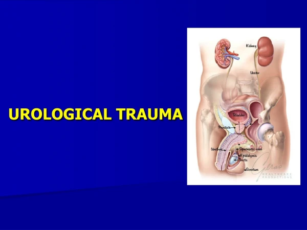
Urological Trauma
470 likes | 905 Views
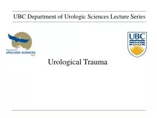
UBC Department of Urologic Sciences Lecture Series. Urological Trauma. Disclaimer: This is a lot of information to cover and we are unlikely to cover it all today These slides are to be utilized for your reference to guide your self study. MCC Objectives.

Urological Trauma
E N D
Presentation Transcript
UBC Department of Urologic Sciences Lecture Series Urological Trauma
Disclaimer: • This is a lot of information to cover and we are unlikely to cover it all today • These slides are to be utilized for your reference to guide your self study
MCC Objectives http://mcc.ca/examinations/objectives-overview/ For LMCC Part 1 Objectives applicable to this lecture: • Urinary Tract Injuries • Kidney • Bladder and Urethra
Objectives Trauma: • Given a patient with a potential urinary tract injury: • To list and interpret key clinical findings • To list and interpret critical investigations • Construct an initial management plan Systems: • Renal • Bladder • Urethra • Ureter • External Genitalia
Case #1 • 55 year old healthy male in MVA, T-boned, high speed • Brought in by ambulance • ABCs done, c-spine cleared • GCS 8 • Presents with gross hematuria • DDx and sites of bleeding?
Case # 1 cont’d • Potential Causes of Hematuria: • Urethral Injury • Bladder Injury • Ureteric Injury • Renal Injury
Renal Trauma Overview • Most commonly injured GU organ • 10% of all serious injuries abdominal have associated renal injury • Variable etiology depending on the area • Rural: 80-95% blunt • Urban: as little as 15% blunt
Hematuria and Renal Injury • NOT related to the degree of injury Gross Hematuria is Variable: • 1/3rd of patients with renovascular injuries • 24% of patients with renal artery occlusion • Only 63% of Grade IV injuries (4% have no hematuria whatsoever!)
Whom to workup • Penetrating trauma: EVERYONE • Blunt trauma: Image with CT if: • gross hematuria • microhematuria plus shock • microhematuria plus acceleration/deceleration Mee et al. (1989) Hardeman et al (1987)
Imaging of trauma patient with hematuria • CT preferred • With contrast • With “delayed” films (mandatory) • Why not get CT cystogram too? • Standard intravenous pyelogram (IVP): Forget it • “One Shot” intraoperative IVP • 2 cc/kg intravenous contrast • Single film at 10 minutes
Intraoperative One Shot IVP • Allows safe avoidance of renal exploration in 32% (Morey et al, 1999) • Highly specific for urinary extravasation • Confirms existence of the other kidney
Indications for renal trauma surgery • Absolute • Grade V renal injury (debatable in blunt trauma): NEPHRECTOMY or REPAIR • Vascular injury in a single kidney: Vascular repair • Relative • Persistent bleeding > 2 units/day • Devitalized segment AND urinary extrav (80% complication rate?) • Renal pelvis injury • Ureter injury • Incomplete staging and ongoing laparotomy • Grade IV vein or artery (thrombosis): nephrectomy • Most penetrating renal injuries
Case • 34 year old man flipped over handlebars of mountain bike • Gross hematuria • Stable • Investigations?
Case • Patient continues to be febrile • Hgb drifts down to 70 after 3 U PRBCs • Management?
Management Options For Renal Trauma • Close observation • Bed rest • Serial Hemoglobins • Antibiotics if urinary extravasation • Radiographic Embolization • Urinary Diversion • Ureteral Stenting • Nephrostomy Drainage • Surgery • Renal Preservation / Reconstruction • Nephrectomy
Bladder: BLUNT: Overview • Rare: <2% of all injuries requiring surgery • Often with a severe associated injuries • Often high-energy injuries • Associated with urethral rupture 10-29% and pelvic fracture 6-10%
Bladder: PENETRATING: Overview • Civilian incidence 2% • Associated major abdominal injuries (35%) and shock (22%) • Mortality high: 12%
Bladder: Diagnosis: Physical Signs • Suspicion: required in cases of penetrating trauma (no time for studies): based on trajectory • Physical signs: • Abdominal pain • Abdominal tenderness • Abdominal bruising • Urethral catheter does not return urine • Delayed? • Fever • No urine output • Peritoneal signs • BUN / Creatinine
Bladder: Diagnosis: Hematuria • Most (95%) have gross hematuria • Microhematuria does occur: usually with minimal injury
Bladder: Diagnosis Plain Cystography • Nearly 100% accurate when done properly: • Adequate filling with 350 cc • Drainage films • Use 30% contrast • Underfilling (250 cc) associated with false negatives
Bladder: Diagnosis CT Cystography • Preferred, especially if already getting other CTs • Antegrade filling by “clamping the Foley” is not OK! • Must dilute contrast (6:1 with saline, or to about 2-4%)
Bladder: Diagnosis CT Cystography Extraperitoneal Intraperitoneal
Posterior Urethra Trauma: Etiology • 4-14% of pelvic fractures • Bilateral pubic rami fractures (straddle fracture) and sacroiliac diasthasis • Mostly males, but can happen in females • Associated bladder rupture in 10-17% • Rectal injury can lead to urethral-rectal fistula in 8%
Posterior Urethra Trauma: Diagnosis • Blood at meatus: 50% • “High riding prostate”: 34% • Inability to urinate • Inability to place urethral catheter • Rarely, perineal hematoma (late finding)
Retrograde Urethrogram Urethral Injury Normal
Posterior Urethra Trauma: Management • Unable to get Foley in: Place an open suprapubic catheter • Allows inspection/repair of the bladder for associated injury • No evidence that s/p “infects orthopedic hardware” although ortho docs worry about it
Testes Trauma • Rare in general • But, in significant scrotal blunt trauma, rupture can be as high as 50% • Bilateral 1.5% • Assaults and sports injuries predominate • Local anesthetic block may improve exam
Case #2 • 34 y.o. male in high velocity MVA presents to ER • GCS 13, ABCs OK • “cannot void” • Tib-fib, Pelvic #, multiple rib #s and pulmonary contusions • Next step?
Main points : Kidney Trauma • Get a CT in everyone with • Gross hematuria • Microhematuria + deceleration or shock • Treat most kidneys nonoperatively • Indications for operation: • Grade V renal injury • Persistent bleeding • Suspected ureter or collecting system injury • Incomplete staging and ALREADY having lap • Isolate the vessels first
Main Points: Bladder Trauma • Get a CT cystogram if pelvic fracture • Most extraperitoneal ruptures can be managed conservatively, • BUT: Consider treating extraperitoneal bladder ruptures OPEN, especially if undergoing lap and DEFINITELY if undergoing pelvic ORIF • Microhematuria (no gross hematuria) usually means no significant injury to bladder
Main Points: Ureter/Urethra • Suspect ureter injuries and you’ll miss them less • If the Foley isn’t draining, it’s probably not in the right place