Download

1 / 19
190 likes | 385 Views
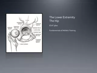
Radiographic Critique of the Lower Extremity. Chapter 5. Foot (AP-Dorsoplantar). ID requirements /marker /No preventable artifacts Contrast & density: soft tissue & bony structures of foot Good penetration trabecular & cortical of phalanges, metatarsals & tarsals (55-65kVp).

E N D
Foot (AP-Dorsoplantar) • ID requirements /marker /No preventable artifacts • Contrast & density: soft tissue & bony structures of foot • Good penetration trabecular & cortical of phalanges, metatarsals & tarsals (55-65kVp)
Foot (AP-dorsoplantar) • ?true AP: 1st, 2nd, & 3rd cuneiforms joints spaces open; with 2cm of calcaneus shown without talar superimposition and equal concavity on both sides of 1st metatarsal midshaft • ?leg, ankle, and foot aligned • ? Rotated laterally the navicular tuberosity is shown in profile with the talus over the calcaneus • ? Rotated medially talus moves away from calcaneus
Foot (AP-dorsoplantar) • ? Tarsometatarsal & navicular-cuneiform joint space open • ?long axis aligned • ? Proximal 2nd & 3rd metatarsal bases in center of field with proximal calcaneus, talar neck, tarsals, metatarsals, phalanges, & surrounding foot soft tissue
Foot (medial or internal)oblique position • Determine ? Obliquity ( 30 or 45 degrees) • Low arch 30 degrees / high arch 45 degrees • ?not rotated enough, 4th & 5th intermetatarsal joint spaces are closed with 4th met base superimposed 5th • ?rotated too much, 4th & 5th intermetatarsal joint space is closed with 5th proximal met base superimposed 4th • ?long axis aligned/ 3rd met base in center of field
Foot (lateral) • ?true lateral: domes of talus are superimposed, the tibiotalar joint is open & distal fibula is superimposed by the posterior half of the distal tibia • ? Long axis of foot at 90 degree angle with lower leg and aligned with long axis • ?proximal metatarsals in center of field with phalanges, metatarsals, tarsals, talus, calcaneus, 1 inch of distal lower leg
Ankle (AP) • If rotated laterally or medially the medial mortise is hidden • ?tibiotalar joint open & tibia seen without foreshortening • ?long axis of tibia aligned • ?tibiotalar joint in center of field with distal 4th of tibia & fibula, talus and soft tissue on film
Ankle (Medial- internal oblique) • ID requirements / marker / no preventable artifacts • Contrast & density with good penetration • ?adequate obliquity: distal fibula seen without talar superimposition, with an open lateral mortise, also lateral & medial malleoli in profile • ?tibiotalar joint open and tibia seen without foreshortening • ?calcaneus seen distal to lateral mortise and fibula • ?long axis of tibia aligned • ?tibiotalar joint in center of field
Ankle ( lateral) • ?true lateral • ?domes of talus superimposed • ?tibiotalar joint open with distal fibula superimposed by the posterior ½ of distal tibia • ?long axis of foot at 90 degree angle with lower leg • ?long axis of tibia aligned • ?tibiotalar joint at center of field showing talus, 1 inch of 5th metatarsal base, soft tissue and distal ¼ of fibula and tibia on film
Knee (AP) • ?femorotibial joint open • ?anterior and posterior condylar margins of tibia superimposed • ?intercondylar eminence and tubercles are seen in profile • ?fibular head seen distal to tibial plateau • ?patella lies superior to patellar surface of femur and is lateral to knee midline • ?intercondylar fossa partially seen • ?femorotibial joint is in center of field with ¼ of distal femur & proximal lower leg with soft tissue on film
Knee (medial & lateral oblique) • ID requirements / marker / no preventable artifacts • Contrast & density with good penetration • 60 to 70 kVp for knee thickness under 5 inches( don’t need grid) • Above 70 kVp for thicker knees for use of grid • ?femorotibial joint open; anterior & posterior condylar margins of tibia are superimposed with fibular head distal to tibial plateau • ?femorotibial joint in center of field with ¼ of distal femur & proximal lower leg on film
Knee (medial oblique) • ?rotated 45 degrees medially • This places the lateral condyle in profile and rotates the fibular head from beneath tibia • Too much-femoral condyles are almost superimposed • Not enough-tibia slightly superimposes the fibular head
Knee (lateral oblique) • ?rotated 45 degrees laterally • This places the medial condyle in profile and rotates the tibia onto the fibula • Not enough-fibular head is seen in center of tibia • Too much – fibular head is aligned with the posterior edge of the tibia and the femoral condyles are almost superimposed
Knee (lateral) • ID requirements / marker / no preventable artifacts • Contrast & density with good penetration • ?knee flexed 10 to 15 degrees with patella proximal to patellar surface of the femur and patellofemoral joint open • ?distal joint surfaces of medial & lateral femoral condyles superimposed with the femorotibial joint space open • ?anterior & posterior surfaces of the medial & lateral femoral condyles superimposed with tibia slighlty superimposing fibular head • ?femorotibial joint in centr of field