Download
1 / 60
690 likes | 1.57k Views
Jugular venous pressure and waveforms. Dr Bijilesh. Jugular venous pulse is the oscillating top of the the distended proximal portion of the internal jugular vein and represents volumetric changes that faithfully reflect the pressure cahnges in the right heart.

E N D
Jugular venous pressure and waveforms Dr Bijilesh
Jugular venous pulse is the oscillating top of the the distended proximal portion of the internal jugular vein and represents volumetric changes that faithfully reflect the pressure cahnges in the right heart
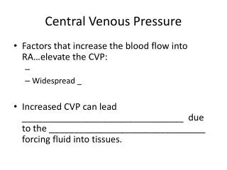
Right atrial pressure during systole and right ventricular filling pressure during diastole • Window into the right heart, providing critical information regarding its hemodynamics.
“In the study of venous pulse we have often the direct means of observing the effects of systole and diastole of right auricle and systole and diastole of right ventricle.” James Mackenzie ..1902
“Precise analysis of the cervical venous pulse and measurement of the height of each wave is not only possible at the bedside but highly desirable” Paulwood ..1950
Anatomy • JV pressure measurement • Causes of elevated JVP • Normal wave pattern • Abnormal wave pattern • Kussmaul s sign and hepatojugular reflux • Specific conditions
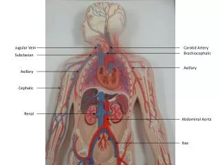
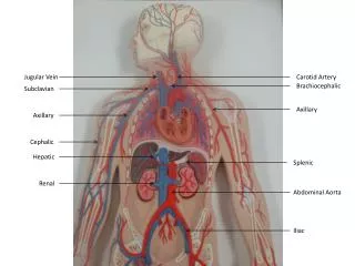
Jugular veins • Internal jugular vein • External jugular vein
Lateral to carotid artery & deep to sternomastoid muscle. • External jugular is superficial to sternomastoid
Examination of JVP • Right IJV is usually assessed both for waveform and estimation of venous pressure • Transmitted pulsations to overlying skin between two heads of sternocleidomastoid
Right IJV Preferred :Why? • Straight line course through innominate vein to the svc and right atrium • Less likely extrinsic compression from other structures in neck Why not EJV • No or less numbers of valves in IJV than EJV
Superficial and lateral in the neck Better seen than felt Has two peaks and two troughs Descents >obvious than crests Digital compression abolishes venous pulse Jugular venous pressure falls during inspiration Abdominal compression elevates jugular pressure Deeper and medial in the neck Better felt than seen Has single upstroke only Upstroke brisker and visible Digital compression has no effect Do not change with respiration Abdominal compression has no effect on carotid pulse Differences between IJV and Carotid pulses
Estimation of Venous Pressure • Measuring jugular venous pressure • Hepatojugular reflux • Examining the veins on the dorsum of the hand • Assessment of jugular venous pressure at bed side reflect mean right atrial pressure
Measurement of JV Pressure • Sternal angle or angle of Louis - reference point • Found approximately 5 cm above the center of the right atrium • Sternal angle – RA Fixed relationship
Position of Patient • Patient should lie comfortably and trunk is inclined by an angle • Elevate chin and slightly rotate head to the left • Neck and trunk should be in same line • When neck muscles are relaxed ,shine the light tangentially over the skin and see pulsations • Simultaneous palpation of the left carotid artery or apical impulse aids in timing of the venous pulsations in cardiac cycle .
Measurement of JVP • Two scale method is commonly used • Normally JV pressure does not exceed 3- 4 cm above the sternal angle • Since RA is approximately 5 cm below the sternal angle , the jugular venous pressure corresponds to 9 cm =7mmhg • Elevated JVP : JVP of >4 cm above sternal angle .
Elevated JVP • Increased RVP and reduced compliance: Pulmonary stenosis Pulmonary hypertension Right ventricular failure RV infarction • RV inflow impedance: Tricuspid stenosis / atresia RA myxoma Constrictive pericarditis
Elevated JVP • Circulatory overload : Renal failure Cirrhosis liver Excessive fluid administration • SVC obstruction
Kussmaul's sign • Mean jugular venous pressure increases during inspiration • Constrictive pericarditis • Severe right heart failure • RV infarction • Restrictive cardiomyopathy • Impaired RV compliance.
Abdominal -Jugular Reflux • Hepatojugular reflux – Rondot (1898) • Apply firm pressure to periumbilical region 30- 60 sec • Normally JV pressure rises transiently to < 1cm while abdominal pressure is continued • If JV pressure remains elevated >1cm until abdominal pressure is continued: Positive AJR.
Abdominal compression forces venous blood into thorax. • A failing/dilated RV not able to receive venous return without rise in mean venous pressure. Positive AJR • Incipient and or compensated RVF • Tricuspid regurgitation • COPD
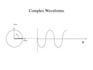
Normal JVP • Normal JVP reflects phasic pressure changes in RA during systole and RV during diastole • Two visible positive waves ( a and v) and two negative troughs ( x and y) • Two additional positive waves can be recorded C wave interrupts x descent and h wave
Normal JVP Waveform • Consists of 3 positive waves • a,c & v • And 3 descents • x, x'(x prime) and y
a Wave • First positive presystolic a wave is due to right atrial contraction • Effective RA contraction is needed for visible a wave • Dominant wave in JVP and larger than v • It precedes upstroke of the carotid pulse and S1, but follow the P wave in ECG
x Descent • Systolic x descent is due to atrial relaxation during atrial diastole • X descent is most prominent motion of normal JVP which begins during systole and ends just before S2 • It is larger than y descent • X descent more prominent during inspiration
C Wave • Not usually visible. • Two different causes - Transmitted carotid artery pulsations. - Upward bulge of closed Tricuspid valve in isovolumic systole
x` Descent • x`descent is systolic trough after c wave • Due to Fall of right atrial pressure during early RV systole • Downward pulling of the TV by contracting right ventricle • Descent of RA floor
v Wave • Begins in late systole ends in early diastole • Rise in RA pressure due to continued RA filling during ventricular systole when tricuspid valve closed • Roughly synchronous with carotid upstroke and corresponds S2 .
y Descent • Diastolic collapse wave (down slope v wave) • It begins and ends during diastole well after S2 • Decline of RA pressure due to RA emptying during early diastole when tricuspid valve opens
h wave • Small brief positive wave following y descent just prior to a wave • Described by Hieschfelder in 1907 • It usually seen when diastole is long • With increasing heart rate, y descent immediately followed by next a wave .
Prominent a Wave • Forceful atrial contraction when there is resistance to RA emptying or increased resistance to ventricular filling • RV inflow obstruction: Tricuspid stenosis or atresia RA mxyoma • Decreased ventricular compliance: • Pulmonary stenosis Pulmonary hypertension of any cause RV infarction RV cardiomyopathy (HOCM) Acute pulmonary embolism
Cannon Waves • Whenever RA contracts against closed TV valve during RV systole • Regular cannon waves: Junctional rhythm VT with 1:1 retrograde conduction Isorhythmic AV dissociation • Irregular cannon waves : Complete heart block Ventricular tachycardia Ventricular pacing or ventricular ectopics .
Absent a Wave • When no effective atrial contraction as in atrial fibrillation
Prominent x descent • Presence of atrial relaxation with intact tricuspid valve and good RV contraction • Causes : Cardiac tamponade Constrictive pericarditis
Reduced x descent • Moderate to severe TR: early sign • Atrialfibriillation
Prominent v wave • Increased RA volume during ventricular systole produce prominent v wave • Severe TR : giant v wave • Giant v wave sometimes causes : systolic movement of ear lobe head bobbing with each systole systolic pulsation of liver pulsatileexophthalmos
PROMINENT V WAVE • ASD with mitral regurgitation • VSD of LV to RA shunt (Gerbode's defect) • RV failure
Rapid y Descent • Severe TR • C .Pericarditis (Friedreich's sign): Early rapid ventricular filling • Severe RV failure • ASD with mitral regurgitation
Slow y Descent • When RA emptying and RV filling are impaired y descent is slow and gradual Tricuspid stenosis Right atrialtumours Pericardial tamponade( y descent may even be absent).
Respiratory influences • Inspiration – increased visibility of venous pulse. Mean venous pressure falls , but the wave forms are accentuated during inspiration.
Waves more prominent during inspiration • X descent more brisk • Increased venous return augment RA contraction and hence relaxation >> brisk x • Also increased venous return augment RV volume and contraction > increased systolic descent of floor of RA>>brisk x’