Download
1 / 26
260 likes | 408 Views
Explore the evolution and functionality of nervous systems in animals, from neurons to brain structure. Discover how neurons work, neural networks, reflexes, and the intriguing behavior of ion channels in nerve cells.

E N D
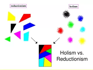
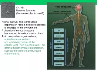
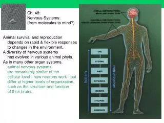
reductionism holism Ch. 48: Nervous Systems: (from molecules to mind?) Animal survival and reproduction depends on rapid & flexible responses to changes in the environment. A diversity of nervous systems has evolved in various animal phyla. As in many other organ systems, animal nervous systems are remarkably similar at the cellular level - how neurons work - but differ at higher levels of organization, such as the structure and function of their brains.
http://www-gpi.physik.uni-karlsruhe.de/pub/robert/Diplom/node5.htmlhttp://www-gpi.physik.uni-karlsruhe.de/pub/robert/Diplom/node5.html In 1911 Ramón y Cajál came up with the idea of the neuron as the basic component of the brain. … a human brain contains some 100 billion ( 1011) neurons with about 1,000 to 10,000 connections each (resulting in 1014 -1015 interconnections). Although one neuron is about 106 times slower than a transistor of a computer the massively parallel processing capability of the brain gives it a much higher efficiency.… it takes about 100-200 msec for the brain to recognize a familiar face … this simple task causes great problems to the computer - if it can do it at all. {efforts at developing AI have been disappointing}
Effector? A Neural Network is an assembly of simple processing elements, whose functionality is loosely based on the neuron. The processing ability of the network is stored in the inter-unit connection strengths, obtained by a process of adaptation to, or learning from, a set of training patterns. {function ‘emerges’ from interactions, that evolve by selection on performance} Functional organization of fundamental (neural) units
recall: herpes emerge from dorsal root ganglia to become shingles Muscle length and velocity are monitored bymuscle-spindle stretch receptors. Activation of these receptors initiates the stretch reflex: motor neurons of antagonists are inhibited (-) & those of synergists are activated (+). {voluntary contraction} Tension on the stretch receptors is maintained by gamma efferent activation of the spindle muscle fibers. {alpha efferent to power effector muscle, gamma efferent (not shown) to spindle keeps sensor stretched as effector shortens} Alpha and gamma motor neurons are often coactivated {in voluntary movement}. http://www.mhhe.com/biosci/ap/vander/student/olc/index.htm + - A simple functional network: the Postural (knee-jerk) reflex;
Ions cannot dissolve in the phospholipid plasma membrane; they must either be pumped by membrane proteins or diffuse through ion channels, which are aqueous pores made of specific transmembrane protein molecules. These molecular channels are selective for specific ions. Membrane potentials are generated by the diffusion of ions and are determined by (a) the ionic concentration differences across the membrane, and (b) the membrane's relative permeabilities to different ions. Plasma membrane Na,K-ATPase pumps maintain intracellular sodium concentration low and potassium high. In almost all resting cells, the plasma membrane is much more permeable to potassium than to sodium;{see gates Fig 48.9} the membrane potential is close to the potassium equilibrium potential, that is, the inside is negative relative to the outside. {about -70mV} http://www.mhhe.com/biosci/ap/vander/student/olc/index.htm All cells have a resting potential (ionic gradients) across plasma membrane, but neurons have voltage-sensitive permeability (voltage-gated ion channels)
Within neurons, graded potentials integrate inputs (like AM, adding analog signals) and action potentials transmit decisions (like FM, w/o degradation) At an inhibitory synapse: an inhibitory postsynaptic potential (IPSP) {hyperpolarization} results when channels to potassium are opened. At an excitatory synapse: an excitatory postsynaptic potential (EPSP) {depolarization} results when channels to sodium are opened. The postsynaptic cell's membrane potential is the result of temporal and spatial summation of the EPSPs and IPSPs at the many excitatory and inhibitory synapses on the cell. {this integrates information; a ‘decision’ at axon hillock} http://www.mhhe.com/biosci/ap/vander/student/olc/index.htm
MS results from autoimmune destruction of myelin An action potential (AP) is a rapid change in the membrane potential during which the potential rapidly depolarizes and repolarizes. {the axon hillock ‘decides’ to fire off an all-or-none AP when local graded potentials integrate to above the threshold, -50mV} APs provide long-distance transmission of information through the nervous system.{along the axon, w/o degradation} http://www.mhhe.com/biosci/ap/vander/student/olc/index.htm APs occur in excitable membranes because these membranes containvoltage-gated sodium channels, which open as the membrane depolarizes,{Fig 48.9} causing a positive feedback toward the sodium equilibrium potential. {Na+ channels open & as Na+ enters, opposing electrical gradient develops} In myelinated nerve fibers, APs manifest saltatory{skippy}conduction. {which allows small diameter axons to be fast; invertebrates have ‘giant axons’} http://www.mhhe.com/biosci/ap/vander/student/olc/index.htm
Most graded signals originate at synapses on the dendritic tree The signal from a pre- to a postsynaptic neuron is a neurotransmitter stored in synaptic vesicles in the presynaptic axon terminal. Depolarization of the axon terminal, which raises the calcium concentration within the terminal, causes the release of neurotransmitter into the synaptic cleft. The neurotransmitter diffuses across the synaptic cleft and binds toreceptors on the postsynaptic cell; the activated receptor usually opens {chemically-gated} ion channels. http://www.mhhe.com/biosci/ap/vander/student/olc/index.htm
http://www.csuchico.edu/psy/BioPsych/neurotransmission.html • Human mood disorders (depressions) are effectively treated with drugs that • block the reuptake of serotonin into the presynaptic axon terminal, • for example fluoxetine (Prozac). Nature419, 872-874 (2002) • Cocaine, opiates, and alcohol produce rewarding effects by • promoting the release or inhibiting the presynaptic re-uptake of dopamine. • {addiction is associated with reduced density of dopamine receptors:} • Parkinson's Disease (PD) is accompanied by a selective destruction of dopamine neurons • in the substantia nigra of the midbrain. PD is treated with L-dopa, a precursor of dopamine in the brain. • Schizophrenia is treated with drugs which block the binding of dopamine to its postsynaptic receptor sites. • Some of the drugs which are used to treat depressive disorders (tricyclic antidepressants) • block the reuptake of monoamines like serotonin at the synapse {and extend it’s effect}. • Endorphins/Enkephalins are endogenous opiates • found in a variety of places (limbic system, midbrain). They are also released as hormones by the pituitary. They are involved in pain reduction and • pleasure (they enhance the effects of dopamine). • Recall NO & Adenosine! • Each time you move a muscle it is because acetylcholine • has been released from a neuron to activate muscle • Alzheimer's Disease is associated with a 90% loss in the brain's • production of acetylcholine in the basal forebrain and hippocampus • GABA (gamma-aminobutyric acid) is the main inhibitory neurotransmitter in the brain. • GLU (glutamate) is the main excitatory neurotransmitter in the brain. • Its actions are mediated at two types of receptor (NMDA and AMPA) involved in memory formation A good source of info: http://www.pharmcentral.com/
The New York Times November 5, 2000HEADLINE:Pesticide Found to Produce Parkinson's Symptoms in Rats.BYLINE: By SANDRA BLAKESLEE … in … Nature Neuroscience. … An organic pesticide widely used on home-grown fruits and vegetables and for killing unwanted fish in the nation's lakes and rivers produces all the classic symptoms of Parkinson's disease in rats Rotenone is extracted from … various tropical plants … Like many plants that produce what are in effect their own pesticides, these plants apparently evolved to produce the compound as a way of warding off insects and other pests. {secondary compounds}Rotenone is found in 680 compounds … organic garden pesticides and flea powders … Parkinson's disease … is caused by the steady loss of cells, in a tiny region of the brain called the substantia nigra, that produce a chemical, dopamine, which is crucial for movement and cognition. … Chronic systemic pesticide exposure reproduces features of Parkinson's disease.Betarbet et al. 2000 Nature Neuroscience 3:1301-1306. The role of environmental agents in Parkinson's diseaseDi Monte DA CLINICAL NEUROSCIENCE RESEARCH 1 (6): 419-426 DEC 2001
Age-adjusted incidence of PD declined consistentlywith increased amounts of coffee intake, from 10.4 per 10000 person-years in men who drank no coffee to 1.9 per 10000 person-years in men who drank at least 28 oz/d. Our findings indicate that higher coffee and caffeine intake is associated witha significantly lower incidence of PD. The data suggest that the mechanism is related to caffeine intake. {Consider correlation vs cause?} Association of coffee and caffeine intakewith the risk of Parkinson disease.Ross GW et al. Journal Of The American Medical Association 283: (20) 2674-2679 MAY 24 2000 Data were analyzed from 30 years of follow-up of 8004 Japanese American men (aged 45-68 years) enrolled in the … Honolulu Heart Program
http://ci.mond.org/9522/952214.html From venoms to toxins to drugs: Toxins have been used scientifically to elucidate physiological mechanisms, as shown by Claude Bernard's classical experiments in the 1850s with curare, the South American arrow poison… the pure alkaloid, tubocurarine, isolated from curare, has been used as a muscle relaxant to accompany general anaesthetic since 1942. {blocks acetylcholine at neuromuscular junction} Another toxin used for its muscle relaxant properties is botulinum toxin, from bacteria Clostridium botulinum The toxin gets into motor nerve terminals and prevents the release of the neurotransmitter acetylcholine … botulinum toxin is often regarded as one of the most toxic substances known … However, its specificity and irreversibility have been useful … When Botox is injected into a muscle, it blocks the nerve impulse from reaching that area, and as a result, the muscle weakens. As the muscle weakens, the skin overlying the muscle relaxes and the wrinkles in the skin gradually soften and often disappear.
Actin-based plasticity in dendritic spinesMatus ASCIENCE 290: (5492) 754-758 OCT 27 2000 Abstract:The central nervous system functions primarily to convert patterns of activity in sensory receptors into …. appropriate behavior. At the anatomical level this requires two complementary processes: a set of genetically encoded rules for building the basic network of connections, and a mechanism for subsequently fine tuning these connections on the basis of experience. … Evidence has accumulated implicating … excitatory synapses made onto dendritic spines, as the sites where connective plasticity occurs. …
Signal-processing machines at the postsynaptic densityKennedy MBSCIENCE 290: (5492) 750-754 OCT 27 2000 Abstract:Dendrites of individual neurons in the vertebrate central nervous system are contacted by thousands of synaptic terminals relaying information about the environment. The postsynaptic membrane at each synaptic terminal is the first place where information is processed as it converges on the dendrite. At the postsynaptic membrane of excitatory synapses,neurotransmitter receptors are attached to large protein "signaling machines" that delicately regulate the strength of synaptic transmission. These machines are visible in the electron microscope and are called the postsynaptic density. By changing synaptic strength in response to neural activity, the postsynaptic density contributes to information processing and the formation of memories. Learning & memory: dynamic complex interactions!
Different classes of postsynaptic glutamate receptors transduce the glutamate signal into electrical & biochemicalevents in the postsynaptic neuron. The -amino-3-hydroxy-5-methyl-4-isoxazolepropionicacid (AMPA)-type glutamate receptor opens {unconditionally} in response to glutamatebinding and mediates most of the rapid excitatory postsynapticcurrent (EPSC). The N-methyl-D-aspartate (NMDA)-type glutamatereceptor … opens {conditionally} in response to glutamateonly when the postsynaptic membrane is concomitantly depolarized. Different patterns of activation of NMDA receptors (NMDARs) canlead to either long-term potentiation (LTP) or long-term depression(LTD) of synaptic strength. These long-lasting forms of synapticplasticity are intensively studied because they may represent ways of encoding "memories" in the brain. • Postsynaptic Signaling and Plasticity Mechanisms • Morgan Sheng & Myung Jong KimScience Oct 25 2002: 776-780. • Excitatory synapses of the brain primarily use glutamate • as their neurotransmitter.
One of PP1's targets is a gene-transcription factor called CREB, which becomes inactive when dephosphorylated by PP1. CREB is required for the synthesis of proteins involved in in memory formation7. These data … suggest that forgetting in ageing may not … result from an irreversible rundown of molecular components but rather from the active intervention of PP1 Not everything that we learn is useful, {except in this class, of course!} so the brain needs a mechanism to prevent itself being burdened by unhelpful details. The molecular details of this mechanism are now being uncovered. Studies of the molecular and cellular foundations of cognitive processes have come of age with the development of techniques that allow genes to be over-expressed, deleted or modified in mice. These altered animals have been studied from a variety of aspects simultaneously by molecular biologists, neurophysiologists and psychologists. The result is the birth of a field that is unravelling the basis of learning, remembering, and now — as a paper in this issue shows — forgetting. On page 970, Genoux and colleagues report that an enzyme known as protein phosphatase 1 (PP1) actively suppresses memories in mice, both during and after a learning exercise. …
The autonomic nervous system innervates cardiac and smooth muscle, glands … Each autonomic pathway consists of a preganglionic neuron with its cell body in the CNS and a postganglionic neuron with its cell body in a ganglion outside the CNS. Excite: Fight or Flight Relax: Rest & Digest The preganglionic neurons in both divisions release acetylcholine; the postganglionic parasympathetic neurons release mainly acetylcholine, and the postganglionic sympathetic neurons release mainly norepinephrine. Effector organs innervated by the autonomic nervous system generally receive dual innervation. http://www.mhhe.com/biosci/ap/vander/student/olc/index.htm
The Brain evolved (phylogeny) & develops (ontogeny) as an elaboration of the dorsal hollow neural tube sensory & motor mapping & ‘cognition’ homeostasis & emotions routing & switching homeostasis & movement visceral homeostasis
“As anyone who has sat through a lecture on a warm spring day knows, attentiveness and mental alertness vary from moment to moment. “ !!! The Reticular Activating System is a functional group of neurons that plays a role in sleep, arousal and attention. Seems to ‘decide’ what needs to be brought to conscious attention, ex: ‘the cocktail party phenomenon’ - hearing your name in a noisy room …
These mappings are variable across people, and ‘plastic’ within people (ex after injury) Motor and sensory body surfaces are topographically mapped onto the cerebral cortex w/ ‘association areas’ nearby. (note cross mapping right-to-left & lateralization of ‘jobs’)
The right hemisphere is stronger at pattern recognition, face recognition, spatial relations, nonverbal ideation, emotional processing in general, music; perception of the relationship between images and the whole context in which they occur. {the ‘holostic hemisphere’} The left hemisphere is most adept at language, math, logic operations, serial sequences of information, detailed, speed-optimized activities, fine visual and auditory details. focused perception. {the ‘reductionist hemisphere’} During an infant’s or child’s brain development, … While working with their hands, most right-handed people use the left hand (right hemisphere) for context or holding and use the right hand (left hemisphere) for fine detailed movement.
http://www.bio.psy.ruhr-uni-bochum.de/asymhumans.htm We use different methods to investigate functional cerebral asymmetries … One of them is the visual half-field technique in which stimuli (faces, words, figures, illusory figures) are presented for 180 ms in the left or the right visual field of a person which fixates a cross in the middle of a computer screen. {180ms is too brief to shift eyes} Due to the pattern of projections within the human visual system, stimuli shown on the right are represented in the left hemisphere and vice versa. If words are shown on the right side (left hemisphere), reaction times are faster and percent correct measures are higher due to the language-dominance of the left hemisphere. The opposite is true for faces and abstract figures (Hausmann and Güntürkün, 1999).
http://www.alumni.berkeley.edu/Alumni/Cal_Monthly/September_2001/Making_up_your_mind.asphttp://www.alumni.berkeley.edu/Alumni/Cal_Monthly/September_2001/Making_up_your_mind.asp Functional MRI of a volunteer committing a list of words to memory. The colored spots represent increased blood flow and brain activity. The circled region is a part of the frontal cortex where activity was higher for words that were later remembered than for words that were later forgotten. The ability to observe which structures participate in specific functions is due to a new technique called functional magnetic resonance imaging, fMRI: provides high resolution, noninvasive reports of neural activity detected by a blood oxygen level dependent signal … This new ability to directly observe brain function {blood flow} opens an array of new opportunities to advance our understanding of brain organization … to mental operations … maps of human brain function.
Amygdala activity related to enhanced memory for pleasant and aversive stimuliHamann et al. 1999 Nature Neurosci 2, 289-293. Pleasant or aversive events are better remembered than neutral events. Using positron emission tomography, {not fMRI} we show that bilateral amygdala activity during memory encoding is correlated with enhanced episodic recognition memory … The human amygdala seems to modulate the strength of conscious memory for events according to emotional importance … Maps of pixels in which individual subject rCBF was significantly correlated with individual-subject episodic-memory enhancement The hippocampus, amygdala, some inner portions of the cortex’s lobes and sections of the thalamus and hypothalamus, form a ring around the brainstem called the limbic system: - interacts with neocortex & generates emotions. {Serotonin is a neurotransmitter arising from emotional stimuli of the limbic system. Prozac is a serotonin uptake blocker.}
This is still controversial