Download

1 / 24
250 likes | 337 Views
Detailed information on the muscles, nerves, veins, and lymphatics of the posterior leg compartment. Includes origins, insertions, actions, and nerve supplies of various muscles. Comprehensive guide for anatomical understanding.

E N D
Posterior Compartment of the Leg Dr. Zeenat Zaidi
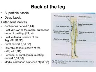
CutaneousNerves • Skin over the popliteal fossa and the upper part of the back of the leg supplied by posterior cutaneous nerve of the thigh • Skin on the upper part of the posterolateral surface of the leg supplied by lateral cutaneous nerve of the calf, a branch of the common peroneal nerve • Skin on the lower part of the posterolateral surface of the leg supplied by sural nerve, a branch of the tibial nerve • Skin on the posteromedial surface of the leg supplied by saphenous nerve, a branch of the femoral nerve
Superficial Veins The small saphenous vein: • Arises from the lateral part of the dorsal venous arch of the foot • Ascendsbehind the lateral malleolusin company with the sural nerve • Follows the lateral border of the tendocalcaneusand then runs up the middle of the back of the leg • Pierces the deep fascia and passes between the two heads of the gastrocnemius muscle in the lower part of the poplitealfossa • Has numerous valves along its course.
Terminates by: • joining the popliteal vein, or • joining the great saphenous vein, or • splitting in two, one division joining the popliteal and the other joining the great saphenous vein • Receives numerous small veins from the back of the leg • Communicates with the: • Deep veins of the foot • Great saphenous vein via anastomotic vessels that run upward and medially
Lymphatics • Lymph vessels run upward and: • Either pass forward around the medial side of the leg to drain into the vertical group of superficial inguinal nodes Or • Drain into the popliteal nodes
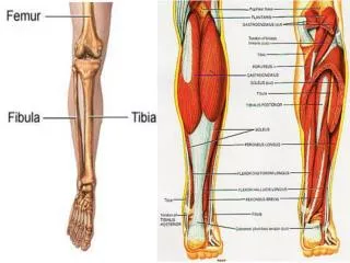
Contents of the Posterior Fascial Compartments of the Leg • Muscles: • The deep fascia of the leg forms a transverse intermuscular septum that divides the muscles of the posterior compartment into superficial and deep groups • Blood supply: Posterior tibial artery • Nerve supply:Tibial nerve
Superficial group of Muscles: Gastrocnemius Plantaris Soleus Triceps surae= soleus + two heads of gastrocnemius. Have common insertion on calcaneus via Achilles tendon (tendocalcaneus) Together, the soleus, gastrocnemius, and plantaris: (1) Act as powerful plantar flexors of the ankle joint (2) Provide the main forward propulsive force in walking and running by using the foot as a lever and raising the heel off the ground.
Gastrocnemius Lateral head may have a sesamoid bone • Origin: • Lateral head: Lateral condyle of femur • Medial head from above medial femoral condyle • Insertion: Posterior surface of calcaneum via Achilles tendon (tendocalcaneus) • Nerve supply:Tibial nerve • Action: • Ankle: Planter flexion • Knee: Assists flexion
Soleus • Broad multipennate muscle • Origin: • Shafts of tibia and fibula • Insertion: Posterior surface of calcaneum via Achilles tendon (tendocalcaneus) • Nerve supply:Tibial nerve • Action: • Ankle: Powerful planter flexion • Provides main propulsive force in walking and running
Plantaris • Origin: • Lateral supracondylar ridge of femur • Insertion: Posterior surface of calcaneum • Nerve supply:Tibial nerve • Action: • Ankle: Planter flexion • Knee: Assists flexion May be absent. Tendon may be used for hand surgery
Deep group of Muscles: Popliteus Flexor digitorumlongus Flexor hellucislongus Tibialis posterior
Tibialis Posterior • Origin: • Posterior surface of shafts of tibia & fibula and interosseous membrane • Insertion:Tuberosity of navicular bone and via slips into sustentaculumtali, cuneiforms, cuboid and bases of 2nd-4th metatarsals • Nerve supply:Tibial nerve • Action: • Planter flexion at ankle joint • Inversion of foot at subtalar and transverse tarsal joints • Supports medial longitudinal arch of foot
Flexor HallucisLongus • Origin: Distal 2/3 of posterior surface of shaft of fibula • Insertion: plantar surface of base of distal phalanx • Nerve supply: Tibial nerve • Action: • Flexes distal phalanx of big toe • Plantar flexion at ankle joint • Supports medial longitudinal arch of foot
Flexor DigitorumLongus • Origin:Posterior surface of shaft of tibia • Insertion: plantar surface of base of 2nd-5th distal phalanges • Nerve supply: Tibial nerve • Action: • Flexion of 2nd-5th toes (PIP/DIP/MP joints) • Assists with foot inversion and plantar flexion • Supports medial and lateral longitudinal arches of foot .
Popliteus • Lies in floor of poplitealfossa. • Origin:from the lateral surface of lateral condyle of femur • Insertion: Posterior surface of shaft of tibia above soleal line • Nerve Supply: Tibial nerve • Action: • Flexes leg at knee joint • Unlocks knee joint by lateral rotation of femur on tibia
The popliteus arises inside the capsule of the knee joint The tendon separates the lateral ligament of the knee joint from the lateral meniscus so that the meniscus is free to move and is less prone (as compared to right meniscus which is fused to right collateral ligament)to get damaged in knee joint injuries
Artery of the Posterior Compartment Posterior Tibial Artery • One of the terminal branches of the popliteal artery • Begins at the level of the lower border of the popliteus muscle • Passes downward deep to the gastrocnemius and soleus and the deep transverse fascia of the leg • Lies on the posterior surface of the tibialis posterior muscle above and on the posterior surface of the tibia below. • In the lower part of the leg the artery is covered only by skin and fascia. • Passes behind the medial malleolus, deep to the flexor retinaculum and • Terminates by dividing into medial and lateral plantar arteries
Venae commitantes of the posterior tibial artery join those of the anterior tibial artery in the popliteal fossa to form the popliteal vein. Branches Peroneal artery Muscular branches are distributed to muscles in the posterior compartment of the leg. Nutrient artery to the tibia Anastomotic branches, which join other arteries around the ankle joint Medial and lateral plantar arteries:
Peroneal artery: • A large artery that arises close to the origin of the posterior tibial artery • Descends behind the fibula, either within the substance of the flexor hallucis longus muscle or posterior to it. • Gives off: • Numerous muscular branches • A nutrient artery to the fibula • A perforating branch pierces the interosseous membrane to reach the lower part of the front of the leg. • Ends by taking part in the anastomosis around the ankle joint.
Nerve of the Posterior Compartment Tibial Nerve • Larger terminal branch of the sciatic nerve • Descends through the popliteal fossa and passes deep to the gastrocnemius and soleus muscles • Lies on the posterior surface of the tibialis posterior and, lower down the leg, on the posterior surface of the tibia • Accompanies the posterior tibial artery and lies at first on its medial side, then crosses posterior to it, and finally lies on its lateral side. • The nerve, with the artery, passes behind the medial malleolus, between the tendons of the flexor digitorum longus and the flexor hallucis longus • Lies deep to the flexor retinaculum and divides into the medial and lateral plantar nerves.
Branches Muscular branches to the soleus, flexor digitorumlongus, flexor hallucislongus, and tibialis posterior Cutaneous: The medial calcaneal branch supplies the skin over the medial surface of the heel Articular branch to the ankle joint Divides into Medial and lateral plantar nerves inferior and posterior to medial malleolus
Clinical notes • Gastrocnemius and Soleus muscle tears • Produce severe localized pain & swelling over the damaged muscle • Ruptured Tendocalcaneus • Common in middle-aged men and frequently occurs in tennis players. • Occurs at its narrowest part, about 2 in. (5 cm) above its insertion • A sudden, sharp pain is felt, with immediate disability. The gastrocnemius and soleus muscles retract proximally, leaving a palpable gap in the tendon. • It is impossible for the patient to actively plantar flex the foot. • The tendon should be sutured as soon as possible and the leg immobilized with the ankle joint plantar flexed and the knee joint flexed. • Plantaris tendon • Rupture is rare • Can be used for autografts in repairing severed flexor tendons to the fingers (like the tendon of the palmarislongus muscle)