Download
1 / 16
160 likes | 343 Views
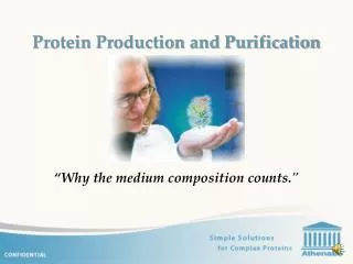
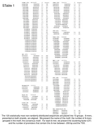
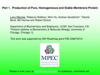
Part 1. Production of Pure, Homogeneous and Stable Membrane Protein. Larry Miercke , Rebecca Robbins, Mimi Ho, Andrew Sandstrom*, Rachel Bond, Bill Harries and Robert Stroud

E N D
Part 1. Production of Pure, Homogeneous and Stable Membrane Protein Larry Miercke, Rebecca Robbins, Mimi Ho, Andrew Sandstrom*, Rachel Bond, Bill Harries and Robert Stroud Department of Biochemistry and Biophysics, UCSF, San Francisco, CA; *Present address is Biochemistry & Molecular Biology, University of Chicago, Chicago, IL This work was supported by NIH Roadmap grant P50 GM073210 Please e-mail Larry at merc@msg.ucsf.edu for questions, comments ect.
Introduction Our approach to growing quality protein crystals is the production of Pure, Homogeneous and Stable Protein. For it is only when identical crystal building blocks are used that the probability of growing large well ordered crystals is optimized. Protein production consists of 4 basic steps: membrane preparation, solubilization, purification & chromatographic characterization, and concentration. Starting with washed membranes, the target is solubilized, then purified to a stable and homogenous state, and finally concentrated while maintaining homogeneity and ultimately minimizing the detergent concentration before entering crystallization trials.
Membrane Preparation: Targets enter the purification workflow once membranes from ≥ 500ml cultures with over-expressed and properly targeted constructs are produced. Most membranes are lysed and washed using a single centrifugation step (harvested cells resuspended directly into high salt buffer prior to lysis, and unwashed pelleted membranes resuspended in glycerol containing buffer). If target proteolysis and protein contaminants are problematic, multiple centrifugation steps are performed (wash cells, lyse in low salt with separate high salt membrane wash; additional steps may include repeated buffer washes and a final sucrose density gradient). A membrane signature gel (series from high to low protein concentration) is run to give an accurate assessment of proteolysis and expression levels, and to determine the membrane concentration required for optimal solubilization screens. Solubilization: Initial solubilization screens consist of three or up to five different detergents (270mM OG, 20mM DDM and 20mM FC12/FC14/MMPC). All properly targeted constructs tested have been found to be solubilized to >80% by at least one of these detergents. Even though OG is generally poor for extraction, it is always included since it is a favorite for crystallization and function. Additional solubilization screens such as Cymals, sterols, POEs, zwittergents, LDAO, TX-100, and diC6PC will be performed next if required for purification. If solubility, homogeneity and stability post affinity purification continue to be problematic, a third approach uses mixtures of detergents, alkyl lipids, and cholesterol. It is not until these all fail to produce quality crystals that selective target extraction studies are implemented.
Purification and characterization: Using a combination of affinity, size exclusion and ion exchange chromatography, six key purification parameters (detergent, pH, ionic strength, reducing agent, osmolytes, and additives) are iteratively examined to find a condition which maintains a PHS concentrated population of protein-detergent-complexes or protein-detergent-lipid-complexes. Affinity chromatography is used for purification and detergent exchange, and size exclusion chromatography is used for purification and for assaying homogeneity. Ion exchange chromatography at low and high pH is used for purification, detergent exchange, assaying solubility and homogeneity dependence on pH and ionic strength, and to help determine if the protein is well behaved (a target which chromatographs on multiple formats indicates a well behaved and identically-folded population). Since oligomerization is the major purification problem, analyzing all generated protein fractions throughout the purification by SEC, including the effects of additional additives and sample storage at 4º and -80ºC, is essential to this purification approach. Concentration: A combination of ultra filtration MWt cut-off filters (centrifugal and high-pressure), chromatography (ion exchange and affinity) and dialysis are all utilized to concentrate protein while minimizing the final detergent concentration. Thin layer chromatography ( is used to estimate the final detergent (and lipid) concentration, while SEC coupled with a tetra detector (absorbance, refraction, viscometer, and light scattering) is used to quantitate the excess micelle concentration while following protein homogeneity and measuring hydrodynamic properties. The focus of this purification approach is quality output, and requires 3-4 days and 7-14 SEC runs per experiment. It is also used following medium throughput expression and purification screens such as 2 detergents for solubilization and SEC (Savage et al 2008), 1 detergent and SEC condition (Min et al 2009), or 1 detergent and 6 different SEC runs (Stroud Lab, unpublished). Over 67 membrane proteins have been purified to PHS using this approach, and 12 crystal structures solved.
Ab Coomassie Membrane Preparation Cells Lyse (high-pressure homogenizer, bead beater) Low speed & high speed spins to access target incorporation (verses IBs) Washes Figure 1. Low speed spin test for membrane incorporation (Zach Newby) BS AS BS AS BS AS Unincorporated Partial Incorporation Complete Incorporation Washed Membranes with properly targeted protein Figure 2. Membrane Signature Gel (Coomassie & Ab, high to low concentration) Accurate assessment of proteolysis and expression levels Determine [membrane] for optimal solubilization screens BEWARE Ab may not bind if [protein] to high Especially for Yeast membranes With Tomomi Tsomeya
Detergent Solubilization Well suspended membranes 300μl Vt, centrifuge tube, stir bar (transfer sup to new tube for mixing) pH 8, 50mM Tris (dictated by first purification step), 300mM NaCl 1 hour, 6 hour, O/N SDS-PAGE Before and After Spin (BS and AS) Round 1 (3-5 detergents) 270mM C8G (OG), 20 mM C12M (DDM) 20mM C14PC (FC14) And 20mM C12PC (FC12) 20mM monoC14PC (MMPC) Round 2 Cymals Sterols POE Zwittergents LDAO TX-100 Round 3, Mixtures of Detergents Alkyl lipids Cholesterol, CHS Under consideration C13M, C14esterS, C14M, C16M Human target Figure 3. BS & AS pairs, R to L OG, 1hr* OG, O/N* DDM, 1hr DDM, 15 hr FC14, 1hr FC14, O/N *Problematic
Detergent Solubilization cont. Figure 4. BS & AS pairs, R to L, of Human target. Top panel = 1hr, bottom panel = O/N 270 mM OG 20 mM DDM 20 mM FC14 20 mM DDM, 3.2 mM CHS 7 mM DDM, 5 mM CHAPS, 3.2 mM CHS 40 mM C12 Sucrose
Detergent Solubilization cont. Figure 5. BS & AS pairs, R to L, of a prokaryotic transporter homolog. Right panel = 15 hr, Left panel = 1 hr(with Tomomi Tsomeya) FC14 SDS DDM MMPC TX100 SDS OG DDM LDAO CHAPS TX100 MMPC Figure 6. BS & AS pairs, R to L, of a human target. 20mM DDM 50mM DDM 100mM DDM 20mM DDM, 3.2 mM CHS 7mM DDM, 5 mM CHAPS, 3.2 mM CHS 20mM FC14 20mM FC14, 20 mM DDM 20mM FC14, 3.2 mM CHS
Purification & Characterization Parameters Detergent/lipid pH Ionic strength Reducing agent Osmolytes Additives Ni/Affinity Desalt Tag Cleavage Cleanup Ni: gravity, 1hr dwell, steps 10DG “desalting” column Thrombin, C3 protease, TEV Optional Co, Ni cleanup Purification Detergent Exchange 10/300mm Superdex 200 0.75/60cm TSK G3000SW ≥ 100mM ionic strength +/- glycerol (5-20% v/v) Purification Homogeneity Size Exclusion Usually start with 10% glycerol 5mM BME/2mM DTT 10DG (3ml/4ml load/elute) Nap 5,10 (0.5ml/1ml, 1ml/1.5ml) Desalt/pH change Purification pH solubility Detergent exchange Well behaved HiTrap 1ml, 5ml SP HP, pH 5-6 Q HP, pH 8-9 Salt gradient, steps Cation and Anion Exchange Common mobile phases 40mM OG, 18mM NG, 8mM DM, 0.5-2mM DDM, 4mM FC12, 0.5mM FC14 Key: assay ALL samples by SE 9-15 SE runs/ pass
Comment on IE: Beware that multi IE peaks may not be real. Since pH and ionic strength are important parameters involved with homogeneity and stability, running S and Q gradients are performed, initially using SEC purified sample. However, as shown in Figure 6 below, different gradients may be needed to verify that multiple peaks are real, or if they are due to a combined effects of [salt] on micelle and detergent belt size/shape thus giving different detergent-protein ratios and shielding charges used in binding. Serial dilutions Figure 7. S/6/OG profiles of identical injections of a human transporter but eluted using different NaCl gradients.
Protein Concentration A combination of ultrafiltration, dialysis and chromatography (Ni, IE, SE) is used. Our most popular MWCO (MWt cut-off) membranes are colored in red. MWCO filters (filter type, method, MWCO) Polyacrylonitrile/polyvinyl chloride (Millipore Amicon XM 50, stir) Favorite; but no longer available (we still have some boxes) Regenerated cellulose Stirred cells Amicon Ultracel YM; 30, 100kDa Tangential spin Amicon Ultracel YM; 30, 50, 100kDa Sartorius (Vivaspin Hydrosart) 30kDa Orbital Biosciences 60, 150kDa Polyethersulfone filters (currently retesting, and testing) Stirred cell Amicon Biomax (PM) 30, 50, 100kDa Pall Omega (modified PES) 30, 50, 100kDa Tangential spin Amicon Biomax (PM) 30, 50, 100kDa Sartorius (Vivaspin PES) 30, 50, 100kDa PALL Omega 30, 50, 100kDa Pressure-fugation Sartorius (Vivaspin, Vivacell; spin +/- pressure; pressure + rock) Dialysis Chromatography (IE, Ni, SE)
Nine Step Expression to Structure Workflow Major Milestones Expression PHS Structure
High Priority MPEC Protein Scorecard (as of 9-08) 1.8 2.0 Å 5.0 12 3.8, 3.5, 5.0 S. cere, HEK, P. past, E. coli, Homologs /E.coli With Corey Anderson, André Bachmann, Sotiri Banakos, Akanksha Bapna, Sarika Chaudhary, Melissa Del Rosario,Vladimir Denic, Robert Edwards, Pascal Egea, Franz Gruswitz, Frank Hays, Joe Ho, David Julius, Monty Krieger, Witek Kwiatkowski, John Lee, Min Li, Bipasha Mukherjee, Vinod Nair, Zach Newby, Roger Nicoll, Sabrina Noel, Joseph O’Connell, Yaneth Robles, Edwin Rodriquez , Zygy Roe-Zurz,Renee Robbins, David Savage, Shimon Schuldiner, Tomomi Tsomeya, Linda Vuong, Jonathan Weismann, and Ronald Yeh. People with italicized names are no longer working with us.
BS AS FT 40mM Ni beads 300mM desalt Cleave, Co Purification summary of euk12TM/DDM expressed in Yeast Solubilization: 100% 20mM DDM, 1hr Purification: 2mM DDM Ni++: 1mg/L; 1hr 0mM Imidazole bind, 40mM wash, 300mM bump Desalt into SE buffer: 20mM Tris 8.0, 150mM NaCl Cleave: 4unit/OD thrombin, O/N, 95-100% Benzamidine: thrombin removal; Co++: uncleaved removal SE/8.0 PHS dilute & conc., 0.3mg/L yield; 5-10mg PHS preps Homogenous in 0.5mM and 1mM DDM with 10% glycerol 30% lower yields due to enhanced highermers consider assaying highermers (3 equal populations) 2mM DDM required without 10% glycerol Gives larger yield due to minimal highermers IE all binds/bumps on S/6/0.5M as single peak; Q/9 good S/6 best; stay above 20mM NaCl Concentration: 50kDa maximum MWCO; 50kDa dialysis does not remove DDM; S/6/0.5M/4mM DDM Pure Homogenous & Stable Well-behaved 0.5 mg/ml First S/6 7.5mg/ml 1 week T1day T47day
Post Ab/OG Reduced Non-reduced human5TM/OG expressed in HEK293s Pure Post SE/OG Solubilization: 100% 50mM OG, 1hr, whole cell Purification (40mM OG) Ab affinity/50mM OG: 10CV wash 200uM peptide bump; 2mg/L yield Non-cleavable tag TSK SE: 7-9mg/8L PHS Binds and bumps on S/6 and Q/9 Optimum stability and function at pH 6 DTT and glycerol not required Concentration: 50kDa max MWt filter Prep 2 Prep 1 Serial dilutions Well behaved S/6 Homogenous & Stable at pH 6.0, 0.3mg/ml SDX overlay Time 0 and 49 days And at 11.4 mg/ml, 1 week
S/5 SE/7.3 proZTM/OG expressed in E.coli Sol Solubilization: 100% 200mM OG, O/N (started with 270mM OG) Purification 40mM OG, 2 mM βME/DTT, 10% glycerol Ni++: 3mg/L yield; 2hr 35mM Imidazole bind, 40mM wash, 300mM bump Desalt into Ni buffer: 20mM Tris 8.0, 500mM NaCl Cleave: TEV, >5x excess, O/N Co++: TEV removal SE/7.3: PHS dilute mg/ml; 2-2.5mg/L yield; >20mg PHS preps IE: all binds and bumps on S/5,6 and Q/7.3, 8, 9 as single peak single pH 7.3 SE peaks using dilute mg/ml from pH 5-8 (highermers at pH 9) Concentration 50kDa maximum YM MWt cut-off Q/7.3/0.3M/40mM OG---8mM excess OG/16.7mg/ml PDC Properties: 113 moles OG/tetramer, globular (IV=0.05), 4.3 nm Rh, 0.17 dn/dc Ni BS AS FT bump desalt cleaved BS AS Well behaved Homogenous & Stable Pure SDX (pH 7.3, 40mM OG) Pooled SEC fractions prior to Q/7.3 16.7 mg/ml post Q/7.3, freeze thawed 6mg/ml post dialysis Minimized [Detergent] Q/7.3 SDX, 7.3, 40mM OG 8mM excess OG micelles RI 280nm 280nm PDC